Canine Pododermatoses Challenge
Skin diseases of the feet and claws in dogs often look very similar and can be frustrating to diagnose. Definitive diagnosis often requires skin scrapings, impression smears, dermatophyte cultures, biopsies, and even broader diagnostic tests for systemic diseases that affect the skin. Clients have a difficult time understanding this and often expect you to recognize a skin disease "on sight." This might be possible for something simple, like a suspected tumor or a broken claw, but when all of the paws are affected, the bigger picture needs to be considered.
The approach veterinary dermatologists take is to consider the examination findings and the signalment and formulate a "short list" of differential diagnoses. In a clinical setting, you would have the benefit of many additional pieces of information, such as physical examination, history, and diagnostic tests-this exercise does not suggest that a diagnosis should ever be made on appearance alone, and it is unlikely that the reader will make a correct diagnosis in all cases.
1. 2-year-old weimaraner: Contact allergic dermatitis

This dog had a pruritic papular eruption present only ventrally. The young age and the ventral distribution along with pruritus suggest such differential diagnoses as contact allergy (irritant is usually painful), sarcoptic mange, atopic dermatitis, or food allergy. The diagnosis was made by confinement and recrudescence on challenge with the offending outdoor plant. Contact allergic dermatitis can be difficult to diagnose because the only effective means of diagnosis is either with patch testing or through confinement and challenge. Both of these can be problematic, as the patches should remain on the animal for 48 hours and confinement is necessary until improvement is noted, which can require many days, especially in chronic cases. Most cases of contact allergic dermatitis occur without a recent environmental change.
2. 9-year-old Doberman pinscher: Superficial necrolytic dermatitis

Other names for superficial necrolytic dermatitis (SND) include necrolytic migratory erythema, metabolic epidermal necrosis, and hepatocutaneous syndrome. The age of this animal and the short duration of the disorder suggest neoplasia, autoimmunity, or a metabolic disorder. It is often difficult clinically to differentiate between SND and epitheliotropic lymphoma. Biopsies and a thorough medical workup would be required for a diagnosis. The dog has liver disease, although pancreatic glucagonomas have also been associated with this syndrome. Unfortunately, the prognosis is grave.

3. 4-year-old English setter: Bacterial furunculosis
This dog has deep staphylococcal pyoderma, which is a very common form of pododermatitis. It can be primary or secondary, and in some cases there are hair or hair follicle keratin foreign bodies as central targets for granuloma formation interdigitally. Demodicosis should always be ruled out by deep skin scrapings; hair plucks; and in some cases where there is extensive fibrosis, through biopsy. A long course of antibiotic therapy, best determined by culture, should be combined with an intense search for an underlying or predisposing cause.

4. 9-year-old mixed-breed spaniel: Dermatophytosis
This older dog has a focal lesion on only one foot. Focal demodicosis is unlikely at this age, but deep scrapings should be done as well as cytologic evaluation. Focal yeast infection may resemble dermatophytosis, but in this case the dog has a fungal infection caused by Trychophyton mentagrophytes that is not generalized. Because the dog is older, if the lesion does not respond rapidly to therapy, a search for an underlying medical disorder should be made. With fungal identification, consider the history and the environment to determine whether rodents are present as they are the natural hosts.

5. 8-month-old collie: Dermatomyositis
In this case, the breed and the age are key in raising this disease to the top of the differential diagnosis list. Lesions are usually alopecic and can be hyperpigmented, occurring on the face, eartips, tailtip, and over bony prominences especially distal from the trunk. Rule-outs include demodicosis and dermatophytosis. Skin biopsy, a thorough history, and electromyelography or muscle biopsy will confirm the diagnosis.

6. 14-month-old Doberman pinscher: Hookworm dermatitis
The young age and the severity of the pruritus should point you in the direction of a parasite. This dog could not be prevented from self-trauma with mechanical devices, bandages, or corticosteroids. The owner also had "creeping eruption." The animal spent considerable time in a garden where the owner consistently dumped the contents of her cat's litter box. In this case, the dog was not positive for hookworms. The migrating larvae were from the cat. Treatment needs to be aggressive and environmental control measures undertaken. Although this disease is unusual today with the parasiticide therapy we use, it can still occur and is problematic.

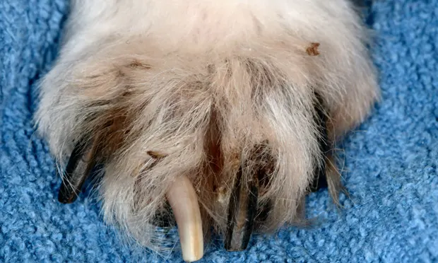
7. 5-year-old German shepherd: Symmetrical lupoid onychodystrophy
Symmetrical lupoid onychodystrophy (SLO) is characterized by spontaneous sloughing or separation of most of the claws, followed by regrowth of misshapen and brittle claws. Onychodystrophy has many differential diagnoses, but this is one of the more common diagnoses when all claws are affected. One method to obtain a definitive diagnosis of claw disease is by p-3 amputation and biopsy. Culture for dermatophytes from claw shaving can help define fungal infection. Secondary bacterial involvement is common and may complicate the condition. The cause of SLO is not known, but some breeds, such as German shepherds, are overrepresented. A variety of treatments have been recommended; the reported results have been mixed.

8. 4-year-old cocker spaniel mix: Malassezia infection
When feet are kept wet by licking, yeast infection often occurs. Dried, brown exudate, which can be scraped off the claws of dogs with pruritic feet, often shows large numbers of yeast organisms when viewed microscopically. This can be secondary to allergy or keratinization disorders, or in rare cases, it can be primary. The disorder is probably secondary in this young mixed-breed dog but should nevertheless be treated aggressively with topical therapy, systemic antifungal treatment, or both.

9. 12-year-old mixed-breed pointer: Epitheliotropic lymphoma (mycosis fungoides)
Neoplasia, autoimmunity, and metabolic disorders should be included on the differential list for an older dog with a brief history of skin disease. Considering the type of lesions, one might also nclude a deep infection of bacterial or fungalorigin depending on the environment of the dog. Biopsy is indicated for diagnosis.

10. 3-year-old West Highland white terrier: Atopic dermatitis
A young terrier that licks his paws should be evaluated for allergic disease. Because the tops of the feet are stained with saliva, contact dermatitis is less likely and food allergy or atopic dermatitis is more likely. A thorough history and detailed physical examination will aid in diagnosis.
11. 11-year-old mixed-breed dog:Mast cell tumor

An older dog with a nodule of short duration is likely to have a mast cell tumor. Solitary nodules on toes usually indicate neoplasia, although they can occasionally represent inflammatory reactions to dermatophytes. In this case, skin biopsy is the diagnostic tool of choice, although aspiration biopsy may be a useful diagnostic tool as well.

12. 18-month-old Doberman pinscher: Demodicosis
The appearance of severe hyperpigmentation on the feet and the suggestion of comedone formation that has been present for 1 year, in addition to the breed, should suggest demodicosis. Deep skin scrapings and hair plucks are indicated and should show many mites in various stages. Cytologic evaluation will also undoubtedly reveal the presence of large numbers of cocci, indicating a concomitant secondary infection. Judging by the appearance of the feet, it is likely that the dog has generalized demodicosis.