A 7-year-old neutered male German shepherd dog was presented for >1 year duration of progressive tenesmus, dyschezia, licking of the anal area, and mucopurulent anal drainage with an odor.
Physical Examination
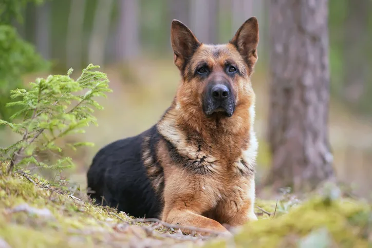
On physical examination, he was reluctant to have his tail lifted. A rectal examination with the patient under sedation revealed mild thickening (2-3 cm) inside the anus. Multiple draining tracts of the perianal tissue encompassed the entire circumference of the anus (Figure 1). The remainder of the examination was unremarkable.

FIGURE 1 Fistulae at the 9-o’clock position (larger) and 4-o’clock position
Ask Yourself ...
What is the appropriate management with the best chance of a successful outcome for this advanced case of perianal fistulae?
A. Surgical management with a laser to ablate each fistula
B. Surgical management with deroofing of each fistula, followed by electrocoagulation or fulguration
C. Administration of prednisone and cephalexin for several weeks
D. Treatment with cyclosporine
Correct answer: D
Treatment Options
Tail amputation was the historical treatment of choice but is no longer recommended. Other surgical options (eg, en bloc resection, laser ± anal sacculectomy) may be incorporated into treatment regimens as adjunctive therapy but have generally been replaced by immunosuppressive or immunomodulatory drugs as the best therapeutic approach.1 Surgical consideration should be reserved for patients with persistent pathology despite extensive attempts at medical management.1 Dogs that experience significant improvement with all lesions except persistent fistulous tracts may require surgical removal of the fistulous tracts.
Administration of immunosuppressive or immunomodulatory drugs, particularly cyclosporine, is the mainstay of medical management. Other immunosuppressive drugs (eg, prednisone, azathioprine, mycophenolate mofetil) can result in complete remission of clinical signs; however, cyclosporine has shown to be effective with the least amount of long-term adverse effects.1-3
Cyclosporine
In a study of 20 German shepherd dogs with perianal fistulae treated with cyclosporine or a placebo, perianal fistulae in all dogs in the cyclosporine group improved at 4 weeks compared with 70% in the placebo group.4 At 16 weeks, 85% of dogs in the cyclosporine group had completely healed; however, lesions recurred in 41% of dogs after discontinuation of cyclosporine; additional cyclosporine treatment was provided or surgical excision was considered in these dogs. Lifelong medical management is thus likely needed because of immune dysregulation and genetics.
Cyclosporine as sole therapy should be started at 7-10 mg/kg PO every 24 hours; however, effective doses can range from 1.5 to 10 mg/kg PO every 24 hours, with a reported increase in efficacy with a higher dose.1 The starting dose can be reduced after complete remission has been achieved. Combining cyclosporine (2-4 mg/kg PO every 24 hours) with ketoconazole (5 mg/kg PO every 24 hours), a competitive binder that decreases cyclosporine clearance by the liver, can increase bioavailability >75%.1,4-6 In practice, the addition of ketoconazole may result in complete remission in 8 to 12 weeks.
Many generic forms of cyclosporine are available for human use, but cyclosporine A is the only form approved for use in animals. Oral generic modified cyclosporine achieves similar bioavailability compared with cyclosporine A7; however, there are rare reports of decreased efficacy with generic modified cyclosporine compared with cyclosporine A. In the authors’ experience, a handful of patients with autoimmune dermatoses (including perianal fistulae) experienced remission with cyclosporine A but relapsed when switched to generic modified cyclosporine. Veterinary formulations have been shown to be bioequivalent.7
Nonmodified and compounded cyclosporine are not recommended due to variable bioavailability and lack of efficacy in controlling the desired disease process.8,9 Research is mixed on whether cyclosporine is best absorbed on an empty stomach or has no effect on bioavilability.7,10 Some dogs cannot tolerate cyclosporine without food due to GI upset. Storage in a freezer may help decrease the incidence of GI upset and does not affect or alter absorption.11,12
Additional Options
Tacrolimus, an immunosuppressive ointment, has a mechanism of action similar to cyclosporine when applied locally.13 Tacrolimus can be used in any treatment protocol as a medication-sparing agent. Topical therapy is usually not recommended as primary treatment for perianal fistulae because of the pain associated with this condition but can be added following complete or partial remission. Topical corticosteroid creams (eg, triamcinolone, nystatin/neomycin/thiostrepton/triamcinolone) can also be used as medication-sparing agents.
Azathioprine (2 mg/kg PO every 24 hours) is an immunosuppressive that functions as a purine analog and disrupts DNA and RNA synthesis, primarily affecting T-cell–mediated immune responses.2 In a study of 14 dogs with perianal fistulae, azathioprine administered in conjunction with prednisone (2 mg/kg PO every 24 hours for 14 days, then tapered to discontinuation) resulted in a 64% complete remission rate.2
Oclacitinib can be used for treatment of perianal fistulae. In a case report, 2 German shepherd dogs treated with oclacitinib (0.88-1.1 mg/kg PO every 12 hours) achieved complete remission, and adequate control was maintained with dose reductions.14
Fluorescent light therapy (typically 2 consecutive treatments once a week for at least 4 weeks) may be beneficial as sole or adjunctive therapy.15
Mesenchymal stem cell therapy is also a potential future treatment option, may be safe and well tolerated, and may have possible future therapeutic value for treatment of cyclosporine-resistant canine perianal fistulae.16
Antimicrobials have historically been used as a part of multimodal medical management but do not provide additional benefits and are no longer recommended.1 Metronidazole has been used in conjunction with other immunomodulatory medications due to anti-inflammatory effects but should not be used for sole management.1
Options for Medical Management of Perianal Fistulae
Option 1
Cyclosporine: 7-10 mg/kg PO every 24 hours for a minimum of 8 weeks
± prednisone: 1 mg/kg PO every 12 hours for 2 weeks, then 0.5 mg/kg PO every 12 hours for 2 weeks, continued tapering weekly
± novel protein diet
Option 2
Cyclosporine: 2-4 mg/kg PO every 24 hours for a minimum of 8 weeks
Ketoconazole: 2.5-10 mg/kg PO every 24 hours for a minimum of 8 weeks
± prednisone: 1 mg/kg PO every 12 hours for 2 weeks, then 0.5 mg/kg PO every 12 hours for 2 weeks, continued tapering weekly
± novel protein diet
Option 3
Azathioprine: 1-2 mg/kg PO every 24 hours for 16 weeks
± prednisone: 1 mg/kg PO every 12 hours for 2 weeks, then 0.5 mg/kg PO every 12 hours for 2 weeks, continued tapering weekly
± novel protein diet
Option 4
Oclacitinib: 0.8-1.2 mg/kg PO every 12 hours
± prednisone: 1 mg/kg PO every 12 hours for 2 weeks, then 0.5 mg/kg PO every 12 hours for 2 weeks, continued tapering weekly
± novel protein diet
Notes
Use of prednisone in place of cyclosporine can reduce treatment cost but is not recommended for long-term management at high doses. Anti-inflammatory doses of glucocorticoids can result in short-term improvement of patient comfort and help the patient be more amenable to topical therapy, especially in severe cases.
Tacrolimus ointment (0.1%) may be added to any treatment option.
Patients should be re-evaluated monthly until fistulae completely resolve. Drug therapy may be adjusted depending on progress but should not be decreased until complete remission is achieved.
If fistulae are still present after 12 to 16 weeks, the medication regimen should be changed. En bloc surgical resection of the remaining lesions may be considered if draining lesions persist despite exhaustive medical management strategies.
Discussion
Breed Predisposition
German shepherd dogs are most commonly affected by perianal fistulae, but this condition is possible in other breeds (eg, beagles, border collies, Australian shepherds, Chesapeake Bay retrievers, Leonbergers, American Staffordshire terriers, Irish setters).1 Low tail carriage has been theorized to cause fecal matter retention and increased local humidity resulting in perianal fistulae development; however, perianal fistulae are prevalent in breeds without low tail carriage, bringing this theory into question.1
Pathophysiology
Immune Dysregulation
Recent literature has highlighted the importance of immunologic alterations in pathogenesis. A predominant systemic CD3+ T cell response and increased mRNA expression of type 1 T-helper cells has been found in lesional skin of patients with perianal fistulae.1,17 This inflammatory response can alter the cytokine profile of the regional skin to a proinflammatory state with increased expression of interleukin-2.1,2 Furthermore, matrix metalloproteinase (eg, MMP-9, MMP-13) expression is increased in lesional skin of affected dogs.1,18
Genetics
A genetic association has been identified with a specific allele encoding the molecules that comprise class II major histocompatibility complex in a population of German shepherd dogs from the United Kingdom and Finland.1 Single nucleotide polymorphisms in the NOD2 gene have resulted in monocyte and macrophage dysfunction that has been associated with inflammatory bowel disease and perianal fistulae, supporting a genetic and autoimmune basis of pathogenesis for German shepherd dogs.1,17
Fecal Dysbiosis
Fecal dysbiosis may be associated with the pathogenesis of this condition.17 A longitudinal study showed changes in microbiologic diversity and shifts in microflora before and after treatment with cyclosporine and ketoconazole.17
Adverse Food Reaction
Adverse food reaction as a contributing pathogenesis is controversial. In a retrospective study of dogs with dermatologic conditions caused by adverse food reactions, 4 out of 130 dogs had perianal fistulae, and all 4 were German shepherd dogs.19 Additional reports of hydrolyzed and novel protein diets as a part of a multimodal treatment plan exist.19,20 Although a diet trial, ideally with a hydrolyzed or novel protein source, can be recommended as a part of the diagnostic and treatment plan, whether treatment can be maintained with diet as sole therapy is unknown.
Take-Home Messages
Perianal fistulae are an immune-mediated and inflammatory condition of the perianal area with rare reports of communication with the rectum.1,21,22 German shepherd dogs are genetically predisposed.
Perianal fistulae are a diagnosis of exclusion and typically occur in dogs 5 to 7 years of age. The major differential diagnoses are anal sacculitis with rupture, neoplasia (eg, squamous cell carcinoma, apocrine gland anal sac adenocarcinoma), and mucocutaneous lupus erythematosus.
Perianal fistulae should be treated medically with immunosuppressive drugs (eg, cyclosporine).
Novel protein diets can be included in a multimodal management plan, but research on benefit of diet as sole treatment is lacking.
Cleaning the perianal area frequently can be beneficial and has been recommended historically but lacks research support. Perianal fistulae result in open wounds, and good hygiene may be helpful; however, frequent cleaning and wiping may not be tolerated due to the painful condition and adds to caregiver burden.
Treatment can be expensive.
Pet owners should be educated that although dogs typically improve with medical management, most relapse if therapy is discontinued. Significant improvement may require 4 to 6 weeks of therapy.
The lowest effective medication dose should be used.
Surgery should be reserved for cases with anal sac involvement or fistulae that do not respond to exhaustive medical management.