Canine Hip Dysplasia Part 2
Canine hip dysplasia (CHD) is a disease of the coxofemoral joint in which laxity of the joint leads to degeneration of articular cartilage and development of osteoarthritis (OA).
Part 1 addressed genetic implications, signalment, pathophysiology, and diagnosis. Part 2 covers surgical and medical protocols, follow-up, and prevention.
Surgical Management
Juvenile Pubic Symphysiodesis (JPS)
JPS is a surgical option for puppies that are younger than 20 weeks of age and have a positive Ortolani’s sign (see What is Ortolani’s Sign?) or a distraction index (DI) of greater than 0.40 on PennHIP radiography(see PennHIP Distraction Index).
JPS encourages premature closure of the pubic symphysis after treatment of the symphysis with monopolar electrocautery.
Assists in maintaining ventroflexion of the pubis and results in better coverage of the femoral heads.
Slows OA or possibly prevents its development in dogs with mild to moderate hip joint laxity.
Dogs with severe laxity (DI >0.70) are less likely to benefit from theprocedure.1
Complications: Juvenile Pubic Symphysiodesis2
Urethral trauma
Diarrhea
Narrowed pelvic canal
What is Ortolani’s Sign?
Ortolani’s sign is a test of hip laxity used to diagnose CHD in dogs. A positive sign is a “snap” as the head of the femur is moved back and forth into the acetabulum after being moved to the acetabular rim (see Figure 1 in Canine Hip Dysplasia Part 1)
When a positive Ortolani’s sign is detected, the angles of reduction and luxation should be measured and recorded. The angle of reduction is an indication of hip joint laxity, whereas the angle of luxation is an indicator of the slope of the dorsal acetabular rim.
These palpation findings, however, need to be confirmed by a comprehensive radiographic evaluation of the hip.
Source: Surgery STAT: Diagnosis and treatment of juvenile canine hip dysplasia. Henry WB Jr. http://veterinarynews.dvm360.com (accessed September 2011).
Triple Pelvic Osteotomy (TPO)
TPO is an option for immature dogs that have no radiographic signs of OA but generally (although not always) are skeletally immature.
With TPO, the pelvis is cut and the ilium and acetabulum rotated externally to improve coverage of the femoral head, ideally to the point that the Ortolani’s sign is negative.3
OA is likely to progress in dogs following this surgery, but lameness and function improve in the operated limbs from recovery to 5 years aftersurgery.4
Outcome is improved if preoperative angle of reduction is less than 45° and angle of subluxation is less than 20°.5
Complications: Triple Pelvic Osteotomy6
Implantation failure prior to bone healing
Pelvic canal stenosis
Sciatic nerve damage
Urethral trauma
Hyperextension of the tarsus
Femoral Head & Neck Excision (FHNE)
FHNE is an option for dogs of any age, although skeletally immature dogs are at greater risk for regrowth and/or surgical revision.
Removal of contact between the femur and acetabulum may improve lameness.
FHNE may be more cost-effective or appropriate for dogs that are not candidates for JPS, TPO, or THA.
Complications: Femoral Head & Neck Excision7,8
Long-term complications in dogs that weigh more than 12 kg:
Decreased range-of-motion
Muscle atrophy
Decreased weight-bearing
Total Hip Arthroplasty (THA)
THA is a salvage procedure for dogs with severe pain in one or both hips.
Dogs that are not candidates include those with:
Orthopedic disease in the same limb
Neurologic disorders (eg, intervertebral disk disease, lumbosacral disease, wobblers syndrome, degenerative myelopathy)
Candidates must be free of infections, such as gingivitis, cystitis, otitis externa, and pyoderma.9
Dogs that develop a distant postoperative infection must be treated with appropriate antibiotics as soon as possible or risk septic loosening of the implants.
Dogs at an increased incidence of infection at the surgical site include those in which the joint has been operated on previously as well as dogs prone to distant infections.9
Cementless implants may decrease the incidence of postoperative aseptic loosening, infection, intrapelvic granulomas, and neuropathy.9-11
At 6 years after surgery, 87% of cementless implants remained intact and functioning well.9,12
Cemented implants do not require extreme reaming of the femoral intramedullary canal.12-15
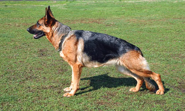
Because of a lack of narrowing of the femoral diaphysis (“stovepipe” femur) and relatively decreased cortical bone width, German shepherds are better candidates for cemented implantation, which is less prone to postoperative subsidence or femoral fracture than cementless implantation is.9,14
Complications: Total Hip Arthroplasty
Aseptic loosening (10%–15% of human cases16)
Femoral fracture (15%–19.5%16-19; more likely in the German shepherd9,14)
Infection (15%–19.5%16-19)
Infarction (15%–19.5%16-19)
Luxation of the hip (up to 20%20-22)
Medical Management
Nonpharmacologic
Diet
Weight reduction lowers the incidence of lameness if an OA patient is overweight.23,24
Keeping dogs with OA slightly underweight may help slow progression and severity of the disease.25,26
Exercise
Exercise can maintain muscle strength, which in turn helps stabilize the joint, slow OA progression, and lessen associated pain.27,28
Controlled exercise prevents excessive force on the joint and articular cartilage.
Swimming or walking on nonslick surfaces (eg, grass lawns, treadmills) is ideal.
Rehabilitation
For dogs with substantial muscle atrophy, physical rehabilitation can be vital in improving lameness through muscle development and may include:
Electrical stimulation
Underwater treadmill therapy
At-home exercises under owner supervision as instructed by a veterinarian with training in rehabilitation
Physical Activities to be Avoided29
Jumping
Running up or down stairs
Leaping
Abrupt stops after running, including catching a thrown ball
Any exercise on slick surfaces
Pharmacologic
Pharmacologic medications are reviewed without respect to their potential for complications; however, when treating patients, the side effects of any treatment must be considered for each patient on an individual basis.
Intraarticular therapy
Stem cell
Fat is taken from the dog surgically; stem cells are then isolated by using special in-house equipment or sending the fat specimen to a processing facility for isolation of the cells, which are then returned and injected into the joint.
Improvement in clinical signs varies and is not permanent; however, improvement can last up to 1 year with very few side effects.30
Botulinum toxin
Dogs receiving intraarticular injection of toxin A reportedly show mild to moderate improvement in lameness and clinical signs.
Improvement is temporary and variable in its effects.31
Hyaluronic acid
Intraarticular injection may decrease matrix metalloproteinases, stimulate chondrocyte proliferation, and decrease degenerative cytokines, such as tumor necrosis factor-a (TNF-a) and interleukin-1.32
Conflicting reports on its effectiveness exist.33-35
Oral therapy
NSAIDs
The cyclooxygenase inhibitors carprofen, meloxicam, and firocoxib have been shown to improve clinical signs of OA in dogs.33,36,37
Side effects include adipsia, anorexia, constipation, diarrhea, emesis, GI ulceration, and polydipsia.37,38
Tramadol
A weak inhibitor of opioid receptors, tramadol interferes with the release and reuptake of noradrenaline and serotonin.
In humans, tramadol combined with an NSAID is effective treatment for OA.
Do not administer tramadol with a tricyclic antidepressant, as doing so may cause serotonin syndrome–producing behavior changes, neuromuscular hyperactivity, and autonomic activation.37,39,40
Amantadine
Amantadine is an N-methyl-d-aspartate (NMDA) receptor inhibitor.41
Coupled with meloxicam, amantadine may provide a greater effect than meloxicam alone for the treatment of OA in dogs.42
Although amantadine is not effective as a primary analgesic following surgery, it may be beneficial if administered with an opioid.37
Eicosapentaenoic acid
Eicosapentaenoic acid can reduce lameness in dogs with cranial cruciate ligament disease.43
Reduced arachidonic acid and prostaglandin E2 in normal joints may occur with eicosapentaenoic acid in certain diets (eg, Purina Veterinary Diets JM Joint Mobility Canine Formula, Hill’s Prescription Diet j/d Canine Mobility, Eukanuba Senior Plus) and in fish oil.43,44
Glucosamine and chondroitin sulfate
Together, these supplements may rebuild damaged cartilage.45
They have no effect on lameness but may reduce matrix metalloproteinases, which can degrade cartilage in the joint.46-48
In combination, glucosamine and chondroitin may restore a more normal synovial fluid environment, which could help slow OA progression.49
Polysulfated glycosaminoglycans (PSGAGs)
PSGAGs (Adequan Canine, adequancanine.us) can protect articular cartilage from degradative enzymes as well as stimulate chondrocytes to produce normal components of articular cartilage.50-53
Puppies (from dysplastic parents) receiving 5.0 mg/kg IM twice a week from 6 weeks to 8 months of age had a reduction in coxofemoral subluxation and improved Norberg angles (measure of hip joint congruity).54
Side effects include inhibited intrinsic clotting and increased buccal mucosal bleeding times.33,36,37,45,55
Green-lipped mussel
Green-lipped mussel has anti-inflammatory properties through tetraenoic acid.
Stabilized lipid preparations may decrease joint swelling and lameness.33,36,56
Elk velvet antler
Improved weight-bearing in arthritic limbs of dogs57
Patient Monitoring
Patients at high risk for developing CHD must be monitored closely the first 5 months of life, with monthly examinations (eg, for Ortolani’s sign) performed by a veterinarian.
Early diagnosis can mean early treatment with either JPS or TPO. Even early medical management can decrease the clinical signs in young dogs and may slow the development of OA and the need for surgery as an adult.
Relative Cost
Surgical management can be costly ($$$$– $$$$$).
Medical management costs over the life of the dog can also add up due to rehabilitation therapy, medical therapy, and therapeutic diets ($$$$).
Prognosis
Because CHD is a chronic disease, prognosis depends on the individual dog’s response to treatment.
Prevention
Early diagnosis in breeds at high risk for CHD may reduce its incidence through controlled breeding programs.
Reducing environmental factors in dogs with CHD may lessen the severity and slow progression of secondary OA.
Future Considerations
Many dogs with CHD and secondary OA can lead comfortable lives as pets with medical management and control of predisposing factors and the environment.
Dogs with severe OA and pain can be managed with FHNE or THA.
After undergoing THA, dogs have been able to return to full function and activity.
Good Breeding ≠ Good Hips
It’s almost frightening how often we deal with CHD in practice. It seems so common that it’s easy to forget how much impact this diagnosis has on families who own these dogs.
Several months ago, I had an owner bring her 5-month-old golden retriever in because of lameness. He is a beautiful dog, with the typical ebullience characteristic of this breed. When I examined his hips, however, I was greeted by an unwelcome and sickening “pop”—in both hips. I had never experienced such a dramatic confirmation of dysplastic hips on physical exam, and I don’t think I covered the horror on my face when it happened. As I looked up at the owner and told her of my suspicions, she completely denied this could be possible. She did agree to radiographs, however.
A few minutes later, I had black-and-white proof that this poor puppy not only suffered from CHD, he essentially had no acetabula. There were no fossae—it was like the femoral heads were just floating in the breeze.
When I showed the owner, her initial look of shock turned to anger: “This is impossible! Both his parents were OFA certified! So what are you telling me, my breeder lied to me?!”
I felt terrible, but I carefully explained that OFA certification does not always guarantee offspring will have normal hips. An appropriate breeding program does not mean that all puppies will be CHD-free. A breeder may be the best in the world, but that does not make his or her puppies immune to risk—a woeful reminder of how CHD is a multifactorial disease.
Certainly my explanation was small consolation for the owner, but it was a lesson for me in how frustrating CHD will be to eradicate and how painful it is for everyone involved.
-- Jennifer L. Schori, VMD
PennHIP Distraction Index
The PennHIP DI is a unique method of quantitatively measuring hip joint laxity. The DI is an indicator of the extent (ie, percent) that the femoral head is displaced from the acetabulum:
Hips with a DI close to 0 are considered tight.
Hips with a DI close to 1 are considered very loose.
A DI of 0.58 means the femoral head is 58% out of the joint. With a DI of 0.75, the head is 75% out of the joint
A hip with a DI of 0.50 is twice as lax as a hip with a DI of 0.25.
Source: What is PennHIP? University of Pennsylvania School of Veterinary Medicine. http://research.vet.upenn.edu (accessed September 2011).