Canine Cryptorchidism
Timmy, a 2-year-old castrated German shepherd dog, was referred to a veterinary internist for evaluation of possible cryptorchidism.
History
Timmy had been adopted as a rescued stray 1 year before presentation. At adoption, routine prescrotal castration was performed for the single scrotal testis present (reportedly the right testis); an extensive abdominal exploratory procedure failed to identify an abdominal testis. According to the owners, Timmy had recently started urine marking and showing interest in female dogs, and he was often distracted during training. Timmy was fed commercial dog food and received monthly flea and heartworm preventive medications.
Related Article: Sexual Development: Male or Female?
Physical Examination
At presentation, Timmy appeared healthy with a body condition score of 3/5. The scrotal sac was empty and atrophied, and the prostate was moderate-sized, bilobed, and firm; palpation did not elicit a painful response.
Laboratory Results & Diagnostics
Results of human chorionic gonadotropin (hCG) response testing completed by the referring veterinarian after consultation with a commercial laboratory revealed a testosterone concentration of 1.7 ng/mL following IV administration of 50 IU/kg hCG (range, intact: 1–7 ng/mL; range, castrated: <0.1 ng/mL). No preadministration samples were submitted by the referring veterinarian.
Related Article: Preoperative Ultrasonography for Retained Testes

Serum luteinizing hormone (LH) concentration, a test not labeled for male dogs, was negative at <1 ng/mL. A single positive test suggests a spayed female, although a second test should be performed at a later date to confirm that the patient has been spayed. Positive tests can occur just before ovulation in intact females.

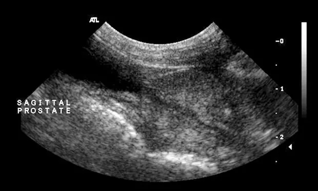
Abdominal ultrasonography—including both inguinal canals to the extent that was possible—disclosed no abnormalities, including presence of an abdominal testis. The prostate appeared enlarged and hyperechoic—typical of a mature male (Figures 1 and 2).
In the presence of a bitch in estrus, Timmy displayed an active libido, and azoospermic semen was readily collected. Semen alkaline phosphatase (SAP) was 26,500 IU/L (>5000 IU/L is indicative of complete ejaculate).
Additional exploratory surgery was performed by a board-certified veterinary surgeon. An abdominal testis was not identified despite thorough evaluation of the abdomen.
Figure 1. Sagittal image of a normal prostate in a castrated dog
Figure 2. Sagittal image of a normal prostate in an intact dog; note the increased echogenicity of the prostatic parenchyma
Why was this dog sent to surgery?
A. Lack of negative feedback (estrogen or testosterone) from the gonads (ovary or testis) to the hypothalamus and pituitary gland results in elevated LH.
B. Prostatic size and echogenicity visualized via ultrasonography supported testosterone influence.
C. Bilaterally cryptorchid dogs are virile (normal testosterone concentrations) but sterile; unilateral cryptorchid dogs with the scrotal testis removed are virile and can be fertile.
D. All of the above
Correct AnswerD. All of the above

On further investigation deep into the left inguinal canal, the left testis was reduced from the canal into the scrotum and removed (Figure 3).
Six weeks postoperatively, the owners reported that Timmy appeared calmer. At follow-up examination, serum LH paired testing was positive (>1 ng/mL).
Timmy’s behavior, prostate size, prostate appearance on ultrasonography, serum testosterone concentration following administration of hCG, and negative LH testing was supportive of endogenous testosterone production.
Spermatogenesis is inhibited by the temperature in the abdomen, and a lack of spermatogenesis was supportive of a nonscrotal testis. Testosterone production is normal in dogs with abdominal testes. The unremarkable ultrasonography results and abdominal exploratory procedures supported an inguinal location of the left testis.
Figure 3. Exteriorized inguinal testis and epididymis; the testis meaused 1.2 x 2.3 cm.
Cryptorchidism
Cryptorchidism, a common congenital genital defect in male dogs, is diagnosed if either or both testes are not present in the scrotum at puberty. Testes typically descend into the scrotum by 6 to 16 weeks of age in puppies and before birth in kittens.
In dogs, descent can occur as late as 10 months of age, but that time frame is abnormal; late testicular descent and undescended testes are heritable defects.
Testicular hormone insulin-like factor 3 is produced by prenatal and postnatal Leydig cells and mediates transabdominal testicular descent from the caudal pole of the kidney to the inguinal canal. This hormone induces growth and differentiation of the gubernaculum from the caudal suspensory ligament.
Transabdominal migration of fetal testes is independent of androgen presence, but inguinoscrotal descent is mediated by testosterone, which causes regression of the cranial suspensory ligament. During the inguinoscrotal phase of migration, shortening of gubernaculum and eversion of cremaster muscle occur.
Localization of one or both cryptorchid testes on ultrasonography can confirm the condition in pediatric patients with unilateral or bilateral involvement and can assist the surgeon in planning the approach (ie, prescrotal vs cranial abdominal).
The Take-Home
Castrated dogs can copulate and ejaculate.
Ultrasonography of the abdomen alone can miss cryptorchid testes in the inguinal canal because of interference from the bony pelvis. Retained testes can be positioned anywhere between the ipsilateral kidney and the scrotum.
In dogs, most SAP is produced in the epididymis. Ejaculate without spermatozoa but with a SAP of >5000 IU/L suggests lack of spermatogenesis.
hCG = human chorionic gonadotropin, LH = luteinizing hormone, SAP = semen alkaline phosphatase
This article is part of the WSAVA Global Edition of Clinician's Brief.
AUTUMN P. DAVIDSON, DVM, MS, DACVIM, is clinical professor in the medicine and epidemiology department at University of California, Davis. She specializes in small animal theriogenology and infectious disease and practices at Pet Care Veterinary Hospital. Dr. Davidson completed her small animal medicine and surgery internship at Texas A&M University and her small animal medicine residency and DVM at University of California, Davis.