Calcium Oxalate Crystalluria
Calcium oxalate (CaOx) crystals can be found in urine samples obtained from healthy dogs and cats as well as from dogs and cats with medical problems. It is important to recognize the crystals themselves, along with confounding diagnostic factors, and to understand what repercussions CaOx crystalluria may have for the patient.
Q: What are CaOx crystals and how do they form?
A: Crystalluria is the presence of microscopic mineral precipitates in urine. The most important variable that influences in vivo crystal formation is supersaturation of the urine with crystallogenic substances, such as calcium and oxalic acid. CaOx crystals can form in acidic, neutral, or alkaline urine.
Q: What is the optimum technique for demonstration of CaOx crystalluria?
A: Samples collected by cystocentesis minimize cellular and bacterial contamination from the urethra and genital tract. To minimize artifacts, evaluate a fresh sample (< 2 hours) at room temperature (crystallization increases with refrigeration and storage). Centrifuge 5 mL of urine at 100 G (approximately 1000 to 2000 rpm depending on the circumference of your centrifuge) for 5 minutes. Gently decant the supernatant and use the remaining liquid to resuspend the sediment. Place a drop on a glass slide, and add a coverslip.
To achieve proper lighting for an unstained sample on a bright-field microscope, close the iris diaphragm and move the light condenser away from the stage. Staining a portion of the sample may help to visualize elements; mix a drop of Sternheimer-Malbin (Clay Adams Sedi-Stain Concentrated Stain, www.bd.com) or 0.5% new methylene blue stain with a drop of the resuspended urine sediment.
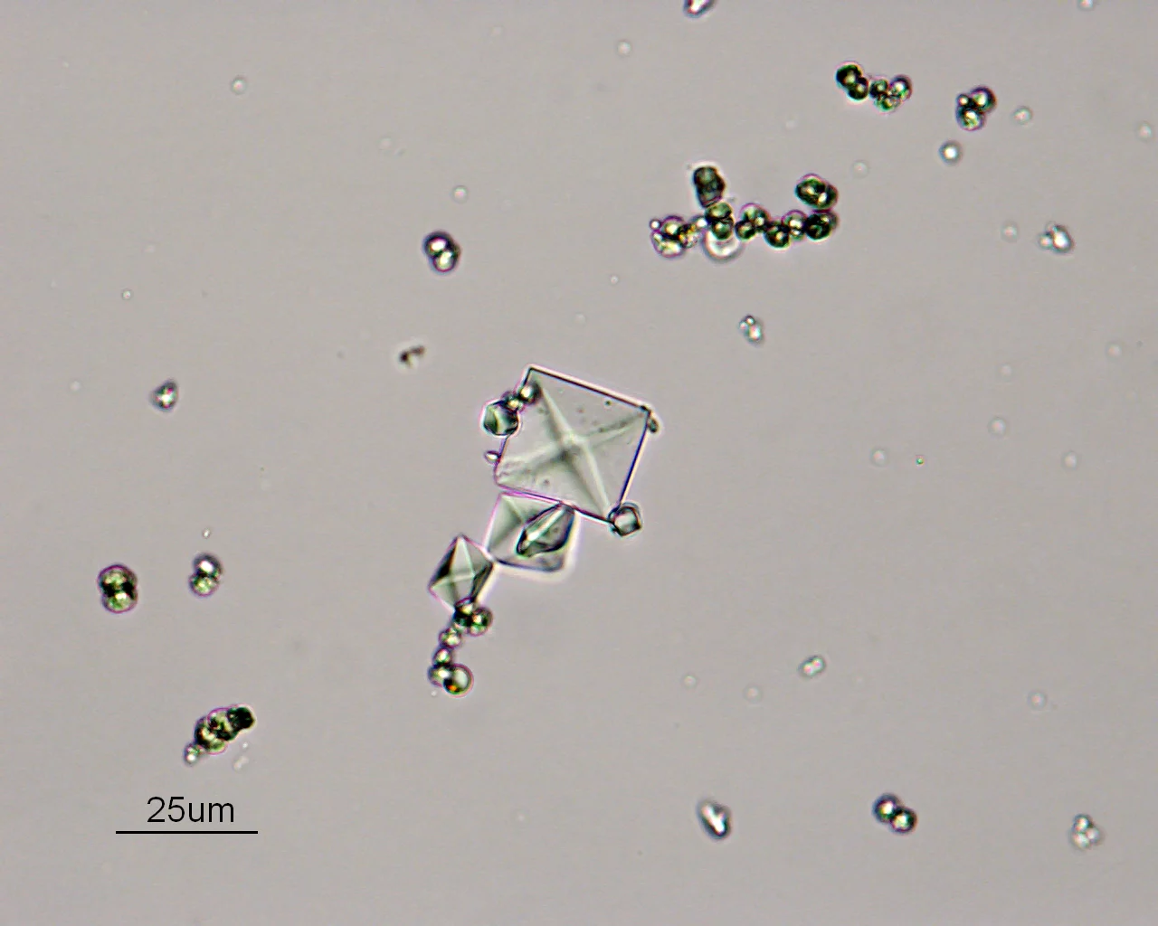
CaOx dihydrate (weddellite) crystals are typically colorless and have a dipyramidal, octahedral, or envelope shape. They often appear as small or large squares whose corners are connected by refractile intersecting diagonal lines (Figures 1 and 2). CaOx monohydrate (whewellite) crystals typically have an elongated, rectangular shape with pointed ends (Figures 3 and 4). They are described as picket fence- or coffin-shaped but may occasionally appear as tiny dumbbells.

CaOx dihydrate crystals. Note the dipyramidal shape and the central set of perpendicular, refractile lines. Small numbers of these crystals may be present in the urine of healthy animals. (Original magnification, 40×)

CaOx dihydrate crystals (Original magnification, 20×)

CaOx monohydrate crystals. Often described as picket fence- or coffin-shaped, these crystals should raise suspicion of ethylene glycol intoxication. (Original magnification, 50×)

CaOx monohydrate crystals (Original magnification, 100×)
Q: What is the relationship between crystalluria and stone formation?
A: Animals with CaOx crystalluria are at increased risk for CaOx urolithiasis; however, CaOx crystalluria should never be interpreted as diagnostic of urolithiasis. Crystals can be found in the urine of animals without stones, and vice versa. Not all animals with crystalluria form uroliths or urethral plugs.
Crystalluria per se does not commonly cause lower urinary tract signs (eg, hematuria, dysuria, stranguria, pollakiuria). Animals with signs of lower urinary tract disease should be carefully evaluated for an underlying cause of the disorder. Urethral plug formation in male cats may be associated with a combination of matrix elements and large aggregates of CaOx crystals, but this is uncommon.
Q: If I find CaOx crystals, what does that mean for my patient? Is treatment indicated?
A: When determining whether CaOx crystalluria is significant, remember that refrigeration promotes formation of CaOx crystals. Some healthy cats and dogs have small numbers of CaOx crystals present in their urine and do not require treatment; however, they should be periodically reevaluated. Excessive CaOx crystalluria or large crystal aggregates in freshly collected urine should prompt examination of the patient for risk factors that result in hypercalciuria or hyperoxaluria.
Hypercalciuria may be caused by hypercalcemia associated with cancer (including lymphoma and various types of carcinoma), primary hyperparathyroidism, hypervitaminosis D, chronic renal failure, and uncommonly osteolytic bone disease. In cats, idiopathic hypercalcemia may result in hypercalciuria and calcium-containing uroliths. However, the underlying cause is not detected in most dogs and cats with hypercalciuria. Hyperoxaluria may be related to dietary oxalate intake (certain plants are rich in oxalate), B vitamin deficiency (reported only in B6-deficient cats), or idiopathic conditions resulting in increased endogenous oxalate production.
If treatment is indicated, increase the patient's water consumption through access to clean water, feeding canned foods, and using drinking fountains (especially for cats) to increased the volume of unconcentrated urine. If dietary manipulation (see next question) and increased water consumption fail to reduce CaOx crystalluria, orally administered potassium citrate supplementation (eg, K-Cit-V Chewable, www.vet-pharmaceutical.com) may be used to try to reduce the magnitude of hypercalciuria and associated CaOx crystalluria. Additional measures might include administration of thiazide diuretics (dogs only) or vitamin B6. Large numbers of CaOx monohydrate crystals are found in patients with a history of exposure to antifreeze and characteristic neurologic and renal signs of such poisoning.
Most of the time, CaOx monohydrate crystals are found in otherwise-healthy animals.
Q: What is the role of diet in decreasing crystalluria?
A: Dietary modifications that are useful in preventing CaOx urolith formation may also be useful in preventing CaOx crystalluria. The diet should be restricted in protein and oxalate content; appropriate commercial diet choices include:
Royal Canin Veterinary Diet Feline Urinary SO
Hill's Prescription Diet u/d (dogs) and x/d (cats)
Eukanuba moderate pH/O Feline
Purina Veterinary Diets UR St/Ox Urinary Feline Formula
The utility of restricting or supplementing dietary sodium chloride is debatable; restriction of dietary phosphorus and magnesium are not indicated.