Brachycephalic Syndrome
Heidi Phillips, VMD, DACVS-SA, University of Illinois
Michelle Jaeger, CVT, VTS (Surgery), University of Illinois
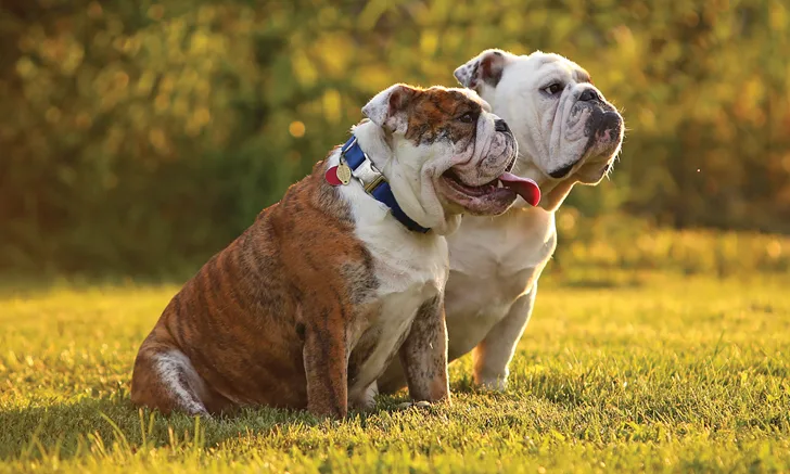
Brachycephalic syndrome affects dogs with markedly shortened craniofacial conformation and can result in dyspnea, exercise intolerance, collapse, and death if not treated appropriately. Corrective airway surgery is best implemented early in life and can be safely performed in dogs as young as 4 to 6 months.1-8 Delaying surgery can predispose brachycephalic dogs to laryngeal collapse, pneumonia, fibrotic lung conditions, and cardiovascular disease, including heart base tumors.7-10 Older dogs may also have more difficulty recovering from surgery.
Case Summary
Heidi Phillips, VMD, DACVS (Small Animal), University of Illinois
Maggie, an 8-year-old spayed English bulldog with a history of noisy breathing, presented to University of Illinois Veterinary Teaching Hospital because of 2 episodes, within 1 week of each other, of severe respiratory distress requiring emergency sedation. An oral examination by her primary veterinarian showed an elongated soft palate, everted laryngeal saccules, and stenotic nares. Maggie was prescribed prednisone for at-home management and referred for surgical assessment and treatment for brachycephalic syndrome.
Brachycephalic syndrome is a chronic, debilitating, and potentially fatal condition in dog breeds with markedly shortened craniofacial conformation.1-3 (See Table 1.)
Brachycephalic Dog Breeds3,16
Congenital and acquired deformities result from selection for craniofacial bony shortening without a concomitant reduction in the volume of nasopharyngeal and oropharyngeal soft tissues.3-8 Anatomic abnormalities include stenotic nares, an elongated soft palate, and a hypoplastic trachea, diagnosed in 58% to 85%, 62% to 100%, and 13% of brachycephalic dogs, respectively.9-11 Aberrant rostral and caudal nasopharyngeal turbinates and intranasal mucosal contact points have also been described.12,13
Most dogs with brachycephalic syndrome have moderate-to-severe upper airway obstruction because of anatomic abnormalities.1 Mild-to-severe respiratory clinical signs predominate and include snoring or stertor, coughing, stridor, inspiratory dyspnea, difficulty eating, exercise intolerance, cyanosis, collapse, syncope, and death.3,7-9 Affected dogs are more likely to experience respiratory signs when stressed or anxious, during exercise or restraint, and in hot weather. Panting caused by overheating can worsen swelling of tissue within the airway. Obesity also contributes to dyspnea in many dogs with brachycephalic syndrome, and an inability to exercise vigorously may predispose these dogs to weight gain.8-9
Most dogs with brachycephalic syndrome have moderate-to-severe upper airway obstruction because of anatomic abnormalities.
Gastrointestinal disorders such as regurgitation, vomiting, gastritis, duodenitis, hiatal hernia, and aspiration pneumonia can accompany respiratory issues.3,10 Brachycephalic dogs may also be diagnosed with sleep disorders, including sleep apnea, which may be linked to systemic hypertension development.3,10 Cardiopulmonary disorders, including hypoxemia, hypercapnea, bronchial collapse, polycythemia, heatstroke, and hypertension, and neoplastic disorders such as chemodectoma may also be present.2,3,7-10,14
Demand for brachycephalic conformation as a breed standard persists despite health issues associated with the anatomic abnormalities.15 The American Kennel Club and Kennel Club of the United Kingdom reported increases of more than 450% in registration of brachycephalic breeds from 2000-2010, indicating a sizable at-risk population.3-4,11,15
Diagnosis
Maggie had a body condition score of 8/9. Physical examination showed stenotic nares. Auscultation of the larynx, trachea, and lung fields disclosed upper airway noise. Routine clinical laboratory tests, including CBC, serum chemistry profile, and urinalysis, were within normal limits. Neck and thoracic radiographs showed a hypoplastic trachea and no signs of underlying cardiac disease or aspiration pneumonia.

Figure 1 Intraoral view of everted tonsils in the brachycephalic patient. Photos courtesy of Heidi Phillips, VMD, DACVS (Small Animal).
Differential diagnoses included brachycephalic syndrome, laryngeal paralysis, upper airway foreign body, neoplasia, and trauma. Following IV administration of butorphanol and propofol, examination of Maggie’s airway disclosed everted, markedly enlarged palatine tonsils (see Figure 1), an elongated soft palate (see Figure 2), and everted laryngeal saccules (see Figure 3). Visual examination of the larynx during inspiration and expiration showed no laryngeal paralysis, but focal contact of the corniculate and cuneiform processes of the arytenoid cartilages on inspiration confirmed mild Stage III laryngeal collapse. The stages are:
Stage I: Everted laryngeal saccules
Stage II: Collapse or paradoxical motion of cuneiform processes of the arytenoid cartilages
Stage III: Collapse or paradoxical motion of the corniculate processes of the arytenoid cartilages
Treatment & Outcome
Treatment
Treatment for laryngeal collapse includes unilateral arytenoid lateralization or permanent tracheostomy, which may predispose patients to aspiration pneumonia or require lifestyle changes, respectively. Because Maggie’s tissue-related airway obstruction was severe, the surgeon chose to treat the airway anatomic abnormalities before addressing the laryngeal collapse.
Maggie was anesthetized, the elongated soft palate was trimmed sharply to the level of the caudal tonsillar crypts, and the oral and nasal mucosa were sutured in a simple continuous pattern of 3-0 poliglecaprone-25. The everted palatine tonsils were trimmed by clamping lateral to the tonsillar tissue with a Kelly hemostat and sharply excising each tonsil with a #15 blade.

Postoperative view of the temporary tracheostomy site. The cranial-most stay suture is passed circumferentially around the tracheal ring just cranial to the tracheotomy and labeled Top. The caudal-most suture is passed circumferentially around the tracheal ring just caudal to the tracheotomy and labeled Bottom. These stay sutures remain in place as long as the tracheostomy tube is needed. They facilitate replacement of a temporary tube removed for cleaning or replacement of a tube inadvertently dislodged.
The everted saccules were removed by gentle grasping of the everted mucosa, pulling toward the midline, and sharply transecting the base of the tissue with a #15 blade. The nares were widened using wedge resection alaplasty. A large pyramid-shaped tissue wedge was resected from the alar wing, and the remaining tissues were joined with interrupted sutures of 4-0 poliglecaprone-25.
Outcome
Postoperatively, Maggie had swelling of the oral and laryngeal tissues, a common consequence of airway surgery, and required temporary tracheostomy during recovery. (See Figure 4.) The tracheostomy tube remained in place while she was monitored and managed in the ICU. The tube was removed on postoperative day 4 after the swelling resolved, and Maggie was able to breathe without significant difficulty.
Team Management
Michelle Jaeger, BS, CVT, VTS (Surgery), University of Illinois
Brachycephalic dogs (see Table 1, in case summary) often experience respiratory difficulties caused by their common anatomic abnormalities. All veterinary team members should be prepared to provide immediate care to brachycephalic patients that arrive in respiratory distress.
Client Communication
Client care team members are usually the first approached about these patients and should instruct a client who calls about a pet in respiratory distress to keep the animal calm and in a cool environment, limit activity, and bring him or her to the practice as quickly as possible for evaluation.
If a brachycephalic patient is in respiratory distress, immediate treatment, including oxygen and sedative administration, and possibly endotracheal intubation or tracheostomy to secure an airway, is required. Also, these patients often become overheated; in these cases, administer IV fluids, apply isopropyl alcohol to the foot pads, or use a fan to cool the patient. If the patient is not in distress but has been brought to the practice because the client is concerned about signs that may be associated with brachycephalic syndrome, team members should take the following steps.
With any brachycephalic patient, a thorough patient history should focus on respiratory issues.
Patient History
With any brachycephalic patient, obtain a patient history that focuses on respiratory issues, including noisy breathing, dyspnea, difficulty sleeping because of open-mouth breathing, coughing, vomiting, regurgitation, gagging, exercise intolerance, heat intolerance, cyanosis, and collapse.1 These signs are caused by anatomical abnormalities (collectively known as brachycephalic syndrome), including stenotic nares, elongated soft palate (see Figure 5), everted laryngeal saccules, hypoplastic trachea, redundant pharyngeal tissue, and laryngeal collapse, which cause upper airway obstruction.2

Elongated soft palate. Photo courtesy of Michelle Jaeger, BS, CVT, VTS (Surgery)
Clear communication with clients during history-taking is important because they often believe their brachycephalic pet’s snoring is normal. The team member should explain that snoring is actually not normal and likely part of brachycephalic syndrome, but proper medical management and surgery could help the pet be more comfortable and prevent a potentially life-threatening situation.
Also, a team member should explain briefly to the client that the veterinarian may suggest various diagnostic tests (eg, thoracic radiography) to evaluate the patient for brachycephalic syndrome and other conditions, such as aspiration pneumonia, hypoplastic trachea, and cardiac disease, and that surgery may be necessary. The client should also be informed the patient may need multiple procedures that will not be identified until the patient is anesthetized and the airway examined.
Patient Evaluation
After studying the patient and the history, the veterinarian should discuss with the client potential surgical procedures that may be necessary following the evaluation, including alar wedge resection, staphylectomy, and laryngeal sacculectomy.
Alar wedge resection involves removing a small piece of the nares to create a wider opening. Staphylectomy entails shortening the soft palate using appropriate anatomic landmarks as a guideline. Often, because of other anatomic abnormalities, the laryngeal saccules become everted and require removal with Allis tissue forceps and Metzenbaum scissors.
The veterinarian should also explain that before any surgery, the patient’s larynx would be examined under light anesthesia to evaluate soft palate length, if the tonsils and laryngeal saccules are everted, and if laryngeal collapse has occurred. Performing this examination and then proceeding immediately with surgery, if necessary, is an advantage because the patient only undergoes one anesthetic event. Treatment with oral steroids to decrease airway swelling may also be indicated.
After the veterinarian’s explanation, a team member should always provide the client with an estimate of all potential charges.
Surgical Procedures
When the decision has been made to perform surgery, the patient should be placed in sternal recumbency with the head resting on a rolled towel. The veterinary nurse should use aseptic technique to prepare the area around the nares and suspend the maxilla using white tape or gauze threaded behind the maxillary canines and tied on either side of the surgical table to IV fluid poles.
If alar wedge resection is performed using either a surgical scalpel blade or carbon dioxide laser, cotton tip applicators and phenylephrine hydrochloride can be utilized topically to control bleeding.3 Long, curved instruments, including Allis tissue forceps, Metzenbaum scissors, and needle drivers, will be used for soft palate resection and everted laryngeal sacculectomy.
Some veterinarians routinely perform tracheostomy following multiple upper airway procedures, whereas others do so only in patients with emergent respiratory distress, but tracheostomy instruments and supplies should always be available when performing any procedure on these patients.
Small-diameter endotracheal tubes should be readily available in case immediate intubation is necessary because brachycephalic dogs often require tubes smaller than other similarly-sized dog breeds that do not have the same anatomical abnormalities.
A brachycephalic patient must be closely monitored postoperatively for respiratory distress.
The veterinary nurse must closely monitor a brachycephalic patient postoperatively for respiratory distress. Instruments needed to reintubate or perform a temporary tracheostomy because of swelling or edema should be readily available. A patient may be placed postoperatively in an oxygen cage for 24-hour monitoring for indications of swelling impacting respiratory function.
Client Care
When the patient is ready for discharge, the veterinary nurse should review at-home care with the client, including:
Elizabethan collar instructions, if an alar wedge resection was performed
Exercise restrictions
Diet restrictions (eg, soft food only for 2 weeks) if staphylectomy or sacculectomy was performed
Recommending harness use when walking
Pain management
Follow-up appointment instructions (eg, when to schedule, if patient should fast before the appointment if suture removal will require sedation)
Long-term management, if necessary (eg, weight loss in obese patients)
Conclusion
Brachycephalic patients require special knowledge and care. Team members should be thoroughly trained and know their role when these patients are brought to the practice, whether their visit is for routine problems associated with their breed, for follow-up care after stabilization, or because they are in respiratory distress.