Brachycephalic Obstructive Airway Syndrome
Sophie Eiger, VMD, University of Florida
W. Alex Fox-Alvarez, DVM, MS, DACVS-SA, Community Care Veterinary Specialists, Gainesville, Florida
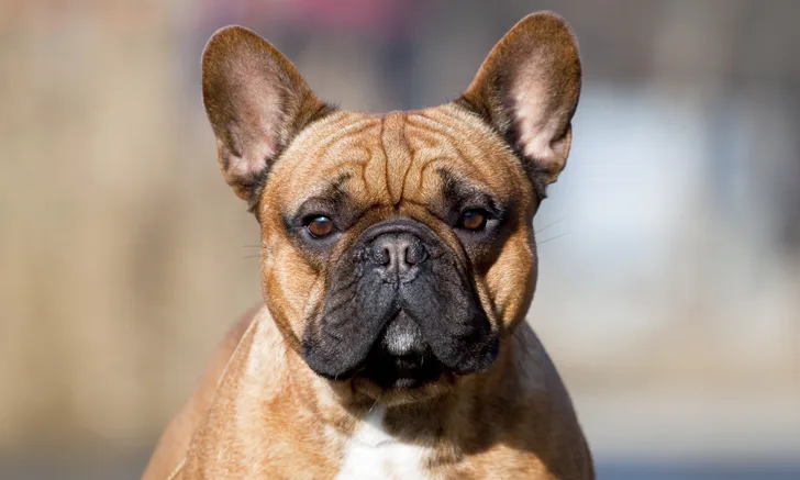
A 2-year-old spayed French bulldog is presented in respiratory distress. The patient was being exercised outside and experienced progressive stertor and dyspnea prior to presentation. She has a history of snoring, exercise intolerance with inspiratory effort, and occasional weekly regurgitation. Brief physical examination reveals moderately stenotic nares and a respiratory rate of 60 breaths per minute with loud upper airway stertor. Heart rate is 160 bpm, temperature is 103.5°F (39.7°C), and mucous membranes are cyanotic. The patient is immediately placed in a cooled oxygen cage for 10 minutes and monitored closely. Once the patient is stable, an intravenous catheter is placed and a sedative is administered. She is then placed back in the oxygen cage. Based on the patient’s breed and history, thoracic radiography and a sedated airway examination are performed.
Thoracic radiographs reveal a mildly hypoplastic trachea and no evidence of aspiration pneumonia (Figure 1). A lateral cervical radiograph shows an elongated, thickened soft palate partially obstructing the laryngopharynx and nasopharynx (Figure 2). Airway examination confirmed the elongated soft palate obstruction and tonsillar eversion (Figure 3). Everted laryngeal saccules are also present. Brachycephalic obstructive airway syndrome (BOAS) is diagnosed. Folded flap palatoplasty, laryngeal sacculectomy, and alarplasty are recommended after improvement in airway swelling associated with the acute episode.

FIGURE 1
Left lateral thoracic radiograph demonstrating mildly hypoplastic trachea (arrow)