Biologic Fracture Management
Brian S. Beale, DVM, DACVS, Gulf Coast Veterinary Specialists, Houston, Texas
Biologic fracture management is a good option for comminuted fractures. Preservation of multiple fracture gaps avoids high interfragmentary strain, favoring bone healing. Conversely, fractures reconstructed to a single fracture gap have high strain, which reduces the stimulus for bone callus formation.
Biologic fracture management is a technique used to optimize the effectiveness of well-documented principles of indirect bone healing.1-7 It can be used with all types of fractures, but is particularly helpful for stabilizing comminuted fractures that are difficult to reconstruct. Preservation of multiple fracture gaps avoids high interfragmentary strain, favoring bone healing (Figure A). Conversely, fractures reconstructed to a single fracture gap have high strain, which reduces the stimulus for bone callus formation. Stabilization of fractures using the principles of biological fracture management is performed with the same type of implant systems used with traditional fracture repair, including externally and internally applied devices. The implant system must be able to support any amount of force placed on the fractured bone during the early healing phase, because the fracture has not been anatomically reconstructed into a solid bony column. Biologic fracture management encourages early callus formation; this early callus shares the load with the implant system, thus reducing the chance of implant failure.
Surgical Approach
The success of biologic fracture management depends on the type of surgical approach. When technically feasible, closed reduction and stabilization is the optimal method of treatment. Open surgical approaches can be either traditional or minimally invasive. The key feature of a minimally invasive approach is preservation of the vascular supply to the comminuted fragments at the fracture site. Small comminuted fragments incorporate quickly into the bony callus if left with a vascular pedicle. Anatomical reduction of small fragments is difficult if vascular supply to the fragment is to remain intact. Disruption of blood supply to the small fragments delays callus formation and bone healing. Other advantages of a minimally invasive approach include shorter intraoperative time, less postoperative pain, and earlier return to function.
The skin and fascia are incised where access is needed to the bone for placement of an implant or to view the fracture site. Muscle dissection should be minimal. Gentle retraction of the muscle bellies overlying the fracture allows visualization of the fracture site (Figure B). The fracture hematoma and fracture fragments should not be disturbed. Adequate exposure is needed only to ensure correct placement of the fixation device.

Features of a minimally invasive surgical approach include low patient morbidity, minimal disruption of blood supply to fracture fragments, rapid bone callus formation, and shortened surgical time.
Method of Fixation
Internal and external implant systems can be used to achieve bone healing using biologic fracture management. Internal devices commonly used include the plate-rod system and interlocking-nail and bone plates. External devices that can be used include casts, splints, linear external fixators, and circular fixators. Other implant systems can be used for biologic fracture management as long as the soft tissue envelope is preserved at the fracture site. Regardless of the implant system used, handling of fracture fragments must be kept to a minimum and the implant system must be strong enough to provide adequate support for the fracture until bone callus begins to form.
Internal Devices
Plate-Rod Systems
The combination of an IM pin and a bone plate has been found to be an ideal implant system for biologic management of comminuted fractures (Figure 1A) in dogs7 and cats. Adding an IM pin to the plate (Figures 1B, C) significantly increases stiffness and the estimated number of cycles to fatigue failure when compared with a plate-only system.


Adding an IM pin to a bone plate has been shown to reduce strain on the plate two-fold and subsequently increase the fatigue life of the plate-rod system 10-fold compared with the plate alone.2 In the canine femur, plate strain is reduced by approximately 19%, 44%, and 61% with the addition of an IM pin occupying 30%, 40%, and 50% of the marrow cavity, respectively.3
The IM pin is applied first. It can be used to assist distraction of the fracture as it engages the distal fragment. The pin provides axial alignment and provides partial stability for application of the plate.
The surgeon must be careful to attain rotational alignment before placing the screws of the bone plate. Only two to three screws are needed in the proximal and distal fragment. Monocortical screws are placed if the screw cannot be directed to avoid the IM pin. Open screw holes are protected by the IM pin and early bone callus, decreasing the chance of plate breakage.
Procedure Pearl
The combination of an IM pin and a bone plate has been found to be an ideal implant system for biologic management of comminuted fractures in dogs and cats.
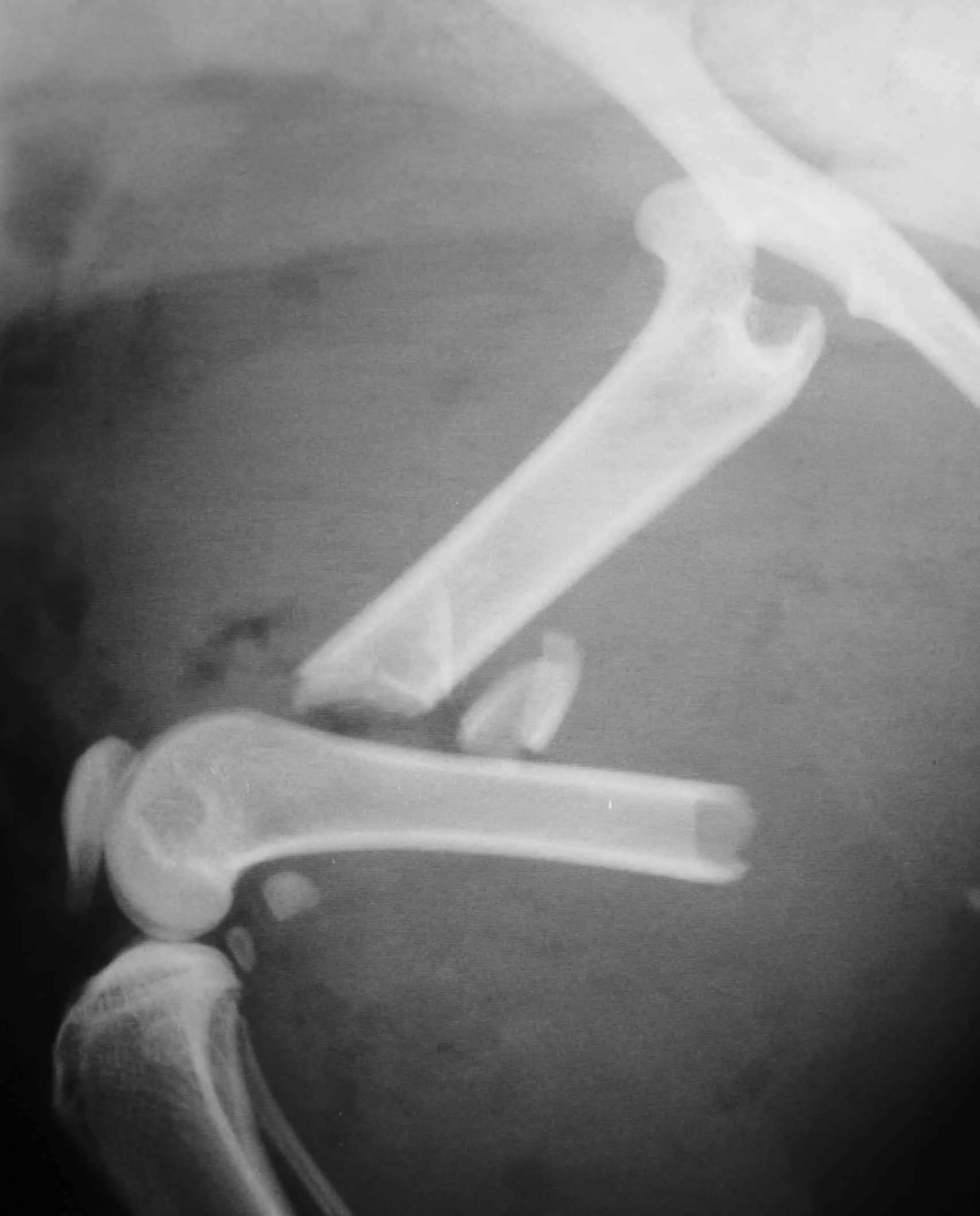
Interlocking-Nail Systems
The interlocking-nail system (Innovative Animal Products, Rochester, MN) is another effective implant system for biologic management of comminuted fractures in dogs and cats.1 The interlocking nail is a modified Steinmann pin having transverse holes designed to accommodate screws or bolts. The addition of screws or bolts increases the ability of the pin to resist rotational and compressive forces at the fracture site (Figures 2A, B). This type of fixation is commonly used for stabilization of fractures of the femur and tibia in humans.


Procedure Pearl
Interlocking nails are easy to apply and are a good option for general practitioners who do not want to invest in a bone-plate system.
Interlocking nails are used in dogs and cats for repair of fractures of the humerus, femur, and tibia. The interlocking-nail system is less expensive than a bone-plate system but has similar biomechanical properties.8 Interlocking nails are easy to apply and are a good option for general practitioners who do not want to invest in a bone-plate system.
Fractures managed using interlocking nails and biologic technique develop extensive bridging callus and early return to function. A minimally invasive surgical approach (note the surgical skin staples) was made to this fracture to minimize disruption of the blood supply to the bone fragment. The fracture fragments quickly become incorporated in the callus if soft tissue attachments can be maintained.

Healing of the comminuted tibial fracture shown in Figures 2A and 2B after stabilization using an interlocking nail system. This fracture reached bony union in 8 weeks.

Postoperative Period and Rehabilitation
A soft-padded bandage can be placed for 2 to 5 days to reduce swelling, decrease pain, and prevent self-trauma to the incision. Prolonged bandaging is generally not necessary unless an external fixator is used.
Early return to weightbearing and light activity is encouraged. Exercise should initially be limited to short, slow walks and range-of-motion exercise.
The duration and intensity of rehabilitation should increase slowly during the time of bony healing. The level of intensity should be adjusted to suit each individual patient.
The goal is to promote range of motion of all the joints of the affected leg and encourage weightbearing at a walk. Ideally, rehabilitation should incorporate strength training and postural and flexibility exercises. Return to full activity is not permitted until radiographic confirmation of bone healing is obtained.
External Fixators
External fixators are often used for biologic management of fractures because of the lower implant cost and relative ease of application. External fixators can be used with closed or open reduction of the fracture. When applied correctly, vascular supply of the fracture fragments can be effectively preserved. Linear external fixators (Figures 3A, B) are most commonly used, but circular fixators are gaining popularity and have advantages for some fracture types. This tibia and fibula fracture achieved bony union in 7 weeks.


Circular fixators are more difficult to apply, and their application is best learned by attending a continuing education short course. The disadvantage to external fixators is the increased postoperative care due to bandage changes and pin management.
Procedure Pearl
Cerclage wires are not used in biologic fracture management due to the increased chance of disturbing blood supplies to fracture fragments.