Epidural Analgesia & Anesthesia
Administration of drugs into the epidural space was first used more than 100 years ago when cocaine was administered epidurally to humans and dogs. With this route of administration, the drug is deposited close to the site of action, maximizing binding to specific receptors and reducing dose-related side effects. Dose requirement of general anesthetics may be reduced, resulting in better systemic homeostasis, and the analgesic effects may last well into the postoperative period.
General Considerations
Epidural administration involves injecting a drug or drug combination into the epidural space—the space between the vertebral canal and dura mater. With spinal administration, the drug is injected into the subarachnoid space, mixing with cerebrospinal fluid.
The term epidural anesthesia describes the injection of a local anesthetic into the epidural space, whereas epidural analgesia refers to the epidural administration of analgesics.When used before surgery, epidural anesthesia or analgesia may reduce the inhalant anesthetic requirement. This is beneficial in all patients but even more so in debilitated and hemodynamically unstable patients. Preemptive analgesia is another important benefit.

Relevant anatomy
In dogs and cats, epidural procedures are usually performed at the lumbosacral space (Figure 1). The spinal cord ends approximately at the level of the sixth and seventh lumbar intervertebral spaces (L6–L7) in medium- to large-breed dogs and at the level of the seventh lumbar and first sacral intervertebral spaces (L7–S1) in cats and smallbreed dogs.Hence, cerebral spinal fluid may be encountered at the level of the lumbosacral space in cats and small-breed dogs.
The tissue planes, arrayed from superficial to deep, at the level of the lumbosacral space are:
Skin
Subcutaneous tissue
Supraspinous ligament
Interspinous ligament
Ligamentum flavum (yellow ligament)
Epidural space.
If the spinal cord is present at that particular level, then following the epidural space are the dura mater, arachnoid, cerebrospinal fluid, pia mater, and spinal cord.The epidural space is not empty; it is filled with variable amounts of adipose and conjunctive tissues and contains blood vessels.
What You Will Need
Required
Clippers
Alcohol and chlorhexidine or povidone-iodine scrubs
Sterile gloves
Spinal needle (Quincke needle; 22- or 20-gauge; 1.5–3.5 inches, depending on patient size)
Optional
3- or 5-mL glass syringe
Sterile water or saline
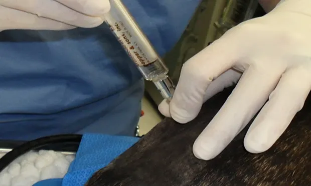
Step by Step: Epidural Analgesia & Anesthesia

Position the patient in sternal or lateral recumbency with the hindlimbs pulled forward. Sternal recumbency facilitates locating the lumbosacral space and using the hanging-drop technique; however, it may worsen fracture sites in the pelvis, femur, or tibia. Thus, lateral recumbency might be preferred in those cases.
Author Insight
To avoid causing more damage to fracture sites while the patient is in sternal recumbency, position the patient close to the edge of the table with the affected leg hanging freely off the side. An assistant should stabilize the patient to prevent it from falling off the table.