Avian Venipuncture
Krystan R. Grant, DVM
Matthew S. Johnston, VMD, Diplomate ABVP (Avian), Colorado State University
Venipuncture is performed for a variety of reasons in avian species, including wellness evaluation, during illness or toxicosis, for DNA testing, and in infectious disease screening.
Wellness examinations should periodically include a complete blood count and a serum biochemical profile. Blood analysis may also help identify abnormalities in debilitated or diseased birds, and DNA testing is used to determine the sex of a bird when, for example, female reproductive problems are suspected.
Successful avian venipuncture is incumbent on knowing the correct needle and syringe sizes to use, the types of samples required for the tests being run, and the amount of blood required. Since requirements may vary between laboratories, it is imperative to contact the reference laboratory to determine submission requirements prior to collecting samples.
A bird’s size determines the needle and syringe size as well as the amount of blood that can be taken safely. A sample should not exceed 1% of the bird’s body weight (eg, sample should not exceed 0.3 mL for a 30-gram bird) and care must be taken with severely debilitated birds, because taking the maximum volume may worsen the patient’s condition.
In general, a 25-gauge needle with a 1-mL or 3-mL syringe may be used with most birds. With very small birds, such as a budgerigar or canary, a 27-gauge with a 1-mL syringe may be more appropriate. For larger birds, such as eagles or geese, a 23- or 22-gauge needle with a 3-mL syringe may be used.
Commonly used anticoagulants greatly alter the morphology of avian blood cells, leukocytes in particular. Blood film should be prepared immediately prior to processing the rest of the sample, and if you are submitting to an outside laboratory, slides should be provided along with the whole blood.
Step-by-Step: Avian Venipuncture
What You Will Need
A 1- or 3-mL syringe with a 5/8- to 1-inch 27-, 25-, 23-, or 22-gauge needle or butterfly catheter
Lavender-topped microtainer containing EDTA for hematologic analysis
Green-topped microtainer containing lithium heparin for serum biochemical profile
Isopropyl alcohol and dry cotton balls

Step 1: Evaluate
Carefully evaluate the bird immediately prior to capture and restraint, and stop if the bird shows signs of distress. For example, sample collection should be delayed or performed with great care in birds exhibiting overt signs of illness, particularly respiratory distress.
Procedure Pearl
Limit stress to the patient by gathering required materials before restraining the bird.
Step 2: Capture & Restrain
Psittacines
Restraint of psittacines is performed carefully to ensure the safety of both the patient and the handler. Using a towel, grasp the bird firmly around the neck, positioning the hand just below the beak to prevent the bird from biting. Use the rest of the towel to loosely wrap the wings to the body to prevent injury from flapping and struggling. Maintain a grip on the neck with one hand and use the other to secure the towel and control the feet. Take care to prevent compressing the body, which could impair the patient’s breathing (A).

Waterfowl & Poultry
Other types of pet birds commonly encountered are waterfowl and poultry. Control of the beak and powerful wings is important, especially in larger patients such as this goose (B).

Raptors
When handling raptors, most injuries to handlers result from contact with their powerful talons. Restrain raptors with quick immobilization of the feet, followed by restraint of the head around the neck as shown with this great horned owl. Many handlers wear welder’s or falconer’s gloves (C).

Step 3: Determine Weight
Depending on the type, size, and condition of the bird, weigh it using a gram scale and secure container (A) or a perch (B).
Procedure Pearl
In the past, blood was collected by clipping a toenail short, but this method is considered inhumane and yields hemolyzed samples.


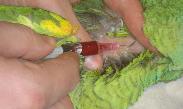
Step 4: Select Vein & Position
The 3 most common sites for venipuncture in birds are the jugular, basilic, and medial metatarsal veins. The right jugular vein, located on the lateral side of the neck, is preferred because, in birds, it is larger than the left vein. The basilic vein often bleeds profusely after puncture and requires a longer period of pressure to achieve hemostasis.
Psittacines
Locate the lateral cervical apterium, a featherless tract of skin near the jugular vein (A). Use alcohol to clear surrounding feathers. The skin is very thin and the vein may be superficial, allowing ready access. It is possible for one person to phlebotomize a small psittacine or a parrot that is under general anesthesia. The head is restrained using the first and middle finger, while the thumb of the same hand is used to hold off the vein near the thoracic inlet (B). Alternatively, an assistant can restrain the body and feet and occlude the vein near the thoracic inlet while the venipuncturist restrains the head and draws the sample.


Larger Birds
The basilic vein, located on the ventral aspect of the elbow, is often used to draw blood from larger birds (C). Restrain the bird in dorsal recumbency with the desired wing extended. Use alcohol to clear the feather area and visualize the vein. The vein may be occluded at the distal humeral aspect. As in smaller birds, the skin is very thin and the vein is superficial.

The medial metatarsal vein, located on the medial aspect of the tarsometatarsus, can also be used in larger birds and is especially useful in waterfowl. The bird is restrained in lateral recumbency, with the desired leg down and extended. The person restraining the bird can occlude the proximal aspect of the vein while the venipuncturist holds the foot (D).

Step 5: Venipuncture
Holding off the vein should allow blood to engorge the vessel, delineating it from the surrounding skin. Gently insert the needle through the skin into the vessel while pulling back on the plunger of the syringe. Hold the syringe and needle steady until the desired amount of blood fills the syringe, as illustrated in basilic venipuncture in a turkey (A) and medial metatarsal venipuncture in a goose (B). Once the desired amount of blood is obtained, apply pressure at the site of venipuncture to avoid a hematoma. Place blood in appropriate container(s) and label them.

