In the Literature
Bohin C, Garcia M, Bertinot C, Graille M, Bernardé A. Compartmental location of middle ear inflammatory polyps in cats: 9 cases (2021-2023). J Small Anim Pract. 2025;66(3):197-202. doi:10.1111/jsap.13811
The Research…
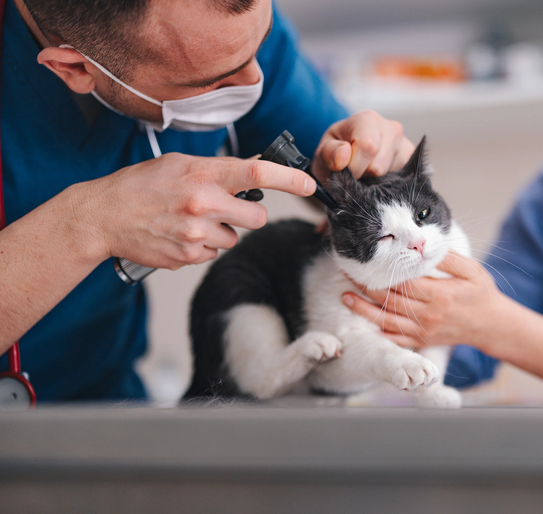
Feline aural inflammatory polyps (AIPs) are nonneoplastic growths of unclear etiology arising from the middle ear or auditory tube and most commonly affect young cats.1,2 Traction avulsion through the external ear canal and ventral bulla osteotomy (VBO) are the main techniques classically described for polyp removal; however, AIP can recur with either of these techniques, with a reported recurrence rate of 0% to 7% after VBO and 0% to 64% after traction avulsion.1,3,4 The feline middle ear contains a nearly complete septum that divides the bulla into dorsolateral and ventromedial compartments; the latter of which is challenging to access using conventional procedures (eg, traction avulsion) via the external ear canal.
This prospective study sought to determine the compartmental location (ventromedial and/or dorsolateral) of AIPs in the middle ear. Client-owned cats (n = 9) with chronic otitis and otoscopic evidence of a pink mass in the deep external ear canal and/or positive CT suggestive of a soft-tissue opacity in the tympanic bulla were enrolled in the study. All cats underwent standard VBO (10 ears; 1 cat had bilateral disease) performed by the same board-certified surgeon that included exploration of the ventromedial compartment which, after breaking down the dividing bulla septum, was followed by exploration of the dorsolateral compartment.
Histopathology confirmed AIP in all 10 ears. AIP tissue filled both compartments in all evaluated cases; solid tissue filled the entire tympanic bulla in 40% of VBOs (with more prominent filling of the ventromedial compartment) compared with 60% solid tissue predominantly in the dorsolateral compartment. The bulla septum was intact in all cases. The exact attachment point of the polyp stalk, including the compartment of origin, could not be determined.
… The Takeaways
Key pearls to put into practice:
AIP tissue tends to occupy both compartments of the feline middle ear. Cats may have a larger volume of tissue in one compartment, suggesting that polyp tissue eventually invades the adjacent compartment despite the point of origin.
The authors concluded that involvement of both compartments provides a possible explanation for the risk of therapeutic failure when using the traction avulsion technique via otoscopy. This technique is limited by the ability to access only the dorsolateral compartment when approaching through the external ear canal. In contrast, the VBO approach allows more complete removal of all macroscopic tissue, attributed to the ability to access both compartments.
Based on anatomic findings of this small cohort, the authors concluded that VBO is necessary for treatment of cats with AIPs. Referral to a board-certified veterinary surgeon may be needed when aural masses are noted on otoscopy.
If surgery for VBO is not viable, chronic tapering of corticosteroids should be considered after traction avulsion to reduce residual polyp tissue. Postprocedural systemic corticosteroids can reduce the rate of recurrence to as low as 0% after traction avulsion.3,4 More studies are needed to corroborate to what degree corticosteroids resolve residual tissue remaining in the inaccessible ventromedial compartment when traction avulsion is used.
You are reading 2-Minute Takeaways, a research summary resource presented by Clinician’s Brief. Clinician’s Brief does not conduct primary research.