Assessing Patient Hydration
Liza Wysong Rudolph, BAS, CVT, VTS (CP-CF, SAIM), East Coast Veterinary Education, Aberdeen, Maryland
Veterinary nurses are often the first member of the veterinary healthcare team to assess patients, so they play a key role in assessing patient hydration. The attending veterinarian prescribes the appropriate intervention, but the veterinary nurse implements the plan and monitors the patient, and he or she must be able to properly assess patient hydration and accurately relay the findings to ensure the patient receives high quality care.
Total Body Water
Healthy animals maintain normal fluid balance in the body (ie, total body water [TBW]) from food and water intake, which compensates for ongoing fluid losses (ie, urination, defecation, respiration). Sick animals may be unable to correct fluid loss through oral intake, which may be exacerbated by vomiting, diarrhea, and polyuria. When total fluid loss exceeds fluid intake, a fluid deficitclinically recognized as dehydrationoccurs.1,2
Total Body Water
An animals lean body weight is approximately 60% total body water (TBW), divided into intracellular fluid (ICF) and extracellular fluid (ECF).
ICF makes up approximately 66% of TBW.
The ICF and ECF are separated by membranes that do not restrict free water movement, and water moves from areas of lesser concentration to greater concentration with ease.
ECF, which makes up the remaining 33% of TBW, is further divided into approximately 25% intravascular fluid (ie, fluid in the blood vessels) and 75% interstitial fluid (ie, fluid present between cells).1-3
The vascular membrane affects the degree of water movement between the intravascular and interstitial space. A deficit in the intravascular space results in hypovolemia and a reduction in perfusion (ie, the transport of blood and oxygen to tissues). A deficit in the interstitial space results in the clinical signs associated with dehydration.
Note that perfusion and dehydration are very closely related but are not synonymous.1
Some conditions can also cause fluid loss into a third space, defined as any body part where large volumes of fluid do not normally collect (eg, pleural or peritoneal space, intestinal or gastric lumen, tissues surrounding trauma sites). Fluid that accumulates in these locations is trapped and not readily available to the body to move back into the cells or vasculature without intervention. This means a patient can have excess fluid retained in the third space but still be dehydrated and hypovolemic, causing potentially serious complications such as edema, reduced cardiac output, and hypotension.1,3
Assessing Patient Hydration
No single factor accurately and easily measures patient hydration. An individualized, multi-faceted assessment of hydration is required that typically includes patient history, physical examination, and laboratory tests.
1. History
Asking unbiased, open-ended questions is an important part of the history and evaluation of hydration status.3 Appropriate questions might include:
How much food and water does your pet normally eat and drink?
Explain any vomiting or diarrhea episodes.
Tell me about any changes you have seen in your pets urination and the timeline of these changes.
What is your pets normal body weight? Have you noticed any changes in your pets weight?
2. Physical Examination
To assess a patients hydration status, examine the following:
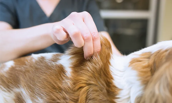
Skin turgor or tenting, which is a measure of how quickly the skin returns to normal after extension, should be tested to help determine interstitial volume.1,4 Gently pull the skin at the back of the neck or along the spine and evaluate how long the skin takes to return to the patients body. A slow return to normal, or decreased skin turgor, indicates a loss of hydration. Conversely, increased skin turgor may be an indicator of overhydration.1,2 This is a subjective assessment, so when possible, test 2 to 3 locations in a single patient. To promote consistency, always use the same locations in each patient.1 Clinical experience is helpful when evaluating a subjective parameter; therefore, practicing subjective evaluation techniques (eg, skin turgor assessment) in many patients is recommended to learn to recognize normal vs abnormal findings. Also, always remember:
Turgor can be falsely increased (ie, a rapid return to normal) by obesity.
Turgor can be falsely decreased (ie, a slow return to normal) by cachexia and in geriatric patients.
Turgor is considered an unreliable hydration assessment in neonates because the high water and fat content in their skin results in increased elasticity.

Normal mucous membrane. Photo courtesy of Erin Layton Photography.
Mucous membranes should be assessed for moisture (eg, moist, tacky, dry).1-3 The oral cavity, specifically the gums, is the most common site for evaluation. Normal mucous membranes should be moist to the touch and shiny in appearance. (See Figure 1.) Tacky mucous membranes have a somewhat sticky quality and are consistent with mild dehydration. Dry mucous membranes, which often develop a dull appearance, indicate a more significant level of dehydration. (See Table 1.) Remember the following important points:
Healthy dogs that pant heavily may have dry gums but be adequately hydrated.
Nauseated patients with ptyalism (ie, hypersalivation) may seem to have moist mucous membranes but in reality may be suffering from dehydration.2
In these cases, alternative sites such as the conjunctiva of the lower eyelid or the mucous membrane lining the prepuce or vulva may be assessed in conjunction with clinical presentation to assess patient hydration.5
Clinical Assessment of Dehydration
*Modified from: Rudloff E. Assessment of hydration. In: Silverstein DC, Hopper K, eds. Small Animal Critical Care Medicine. 2nd ed. St. Louis, MO: Elsevier; 2015:307-310.
Eyes should be visually assessed for the degree of enophthalmos (ie, eyes sunken into the bony orbit) and corneal moisture.1-3
Body weight is an important parameter that should be evaluated in every patient. An acute loss in body weight often can be attributed to loss of body water,1 so an accurate body weight should be obtained on admission and then at least dailyor more often if clinically indicatedto appropriately monitor and evaluate fluid therapy effectiveness in a hospitalized patient.1-3
3. Laboratory Tests
Packed cell volume (PCV) and total solids (TS) may be used to help assess hydration and can be simply and quickly measured with a centrifuged hematocrit tube.2,3,5 The blood sample separates the erythrocytes (ie, red blood cells), buffy coat, and plasma. The percentage of erythrocytes in the sample or PCV is measured with a simple chart. The plasma portion of the sample can be used to obtain a TS, which is measured using a refractometer. The tube is carefully broken, a drop of plasma placed on the refractometer glass plate, and the cover closed. The value is obtained by looking through the eyepiece and reading the g/dL scale corresponding to the level of the horizontal line created by the plasma.
However, always consider that PCV and TS may be affected by multiple factors. For example:
Dehydration often causes a relative increase in PCV and TS because of a TBW deficit (ie, hemoconcentration). Hemoconcentration may falsely increase the PCV in an anemic patient; therefore, a normal PCV and increased TS is consistent with an anemic, dehydrated patient.
Although a decrease in PCV and TS is consistent with hemorrhage or blood loss, an increased or normal PCV with a decreased TS can also be consistent with acute blood loss coupled with splenic contraction, which occurs when the stored erythrocytes are released into circulation in response to acute hemorrhage.
Urine specific gravity (USG) can also help assess hydration. Because USG reflects the urine concentration, an increased USG is consistent with dehydration in patients with normal renal function. A patient with compromised renal function will be unable to properly concentrate its urine and will have inappropriately diluted urine even in the face of dehydration. In this case, assess other parameters that evaluate hydration to properly evaluate the patient.1,2
Conclusion
Veterinary nurses frequently work with patients suspected of dehydration and should be familiar with the signs, as well as the assessments and measurements that should be used to determine the degree of dehydration. Many assessments are subjective, so considering multiple assessments together will help determine a patients hydration status. Veterinary nurses should also be able to identify fluid overload (seeTable 2) in patients receiving fluid therapy.
Signs of Overhydration in a Hospitalized Patient
*Modified from: Donohoe C. Fluid therapy. In: Battaglia A, Steele A. Small Animal Emergency and Critical Care for Veterinary Technicians. 3rd ed. St. Louis, MO: Elsevier; 2016:61-77.
This article originally appeared in the August 2016 issue of Veterinary Team Brief.