Artery Laceration During Cystocentesis
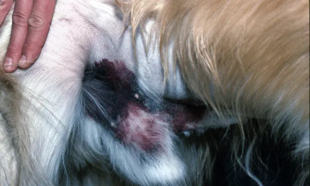
This case illustrates that nothing is routine. A young adult castrated male golden retriever was examined by the neurology service for the primary complaint of weakness. Preanesthetic medical workup included urinalysis of a sample that was collected by cystocentesis. During the procedure the dog became excited and tried to get up. Shortly thereafter, it collapsed from hypovolemia due to substantial bleeding from the cystocentesis site. The dog received 1 whole blood volume (90 ml/kg) of intravenous lactated Ringer's solution under close monitoring and responded well. A pressure bandage was also applied.

Subsequent coagulation test results were normal. The hemorrhage was attributed to laceration of the caudal epigastric artery at the site of the cystocentesis needle stick, which could have been prevented by simply releasing the syringe when the dog moved and tightened his abdominal muscles during the procedure. The illustration shows the contusion and a lateral abdominal radiograph shows a thickened ventral caudal abdominal wall at the site of hemorrhage (arrow).