Anthroscopy in Small Animals
Although descriptions of the use of arthroscopy in small animals date back to the mid 1970s, clinical use of arthroscopy as a primary diagnostic and treatment modality gained popularity in the mid 1990s. This recent expansion of interest results from several factors, including formal training mechanisms, the advent and availability of video arthroscopes and instrumentation specifically suited for small animal procedures, and advanced capabilities for improved treatment in our patients.
AdvantagesThe primary advantage of arthroscopy is its minimally invasive nature, which translates into less tissue trauma, less pain, shorter recovery times, lower complication rates, and more functional recovery than open arthrotomy. The other advantages are less tangible but can be as important; they include visual magnification of the areas of interest, improved access to intraarticular structures, and maintenance of anatomic relationships during surgery. All of these factors lead to more thorough and detailed evaluation of joints and the potential for more precise and optimal treatment.
DisadvantagesThe primary deterrent to use of arthroscopy in small animals is the cost of the equipment, both initially and for maintenance. The smaller scopes and instrumentation used for small animal joints are more fragile and susceptible to damage. Another major reason why orthopedic surgeons are reluctant to incorporate arthroscopy into everyday use is the initial technical challenge. Many hours of formal training and clinical practice are required to gain the proficiency needed to make arthroscopy efficient, atraumatic, and beneficial to patients.

IndicationsThe joints that most commonly undergo arthroscopic evaluation are the stifle, elbow, shoulder, hip, hock, and carpus.
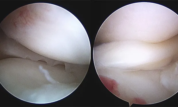
Stifle JointIn our practices, the stifle is the most commonly "scoped" joint. Typical indications include diagnosis and treatment of cruciate ligament disease (Figure 1), meniscal injury (Figures 2A and B), and osteochondritis dissecans (OCD). Arthroscopy provides magnification, access, and fluid distention, allowing a more thorough evaluation of the stifle joint than with

arthrotomy. Damaged cranial cruciate ligaments can be accurately debrided with small power-driven shavers. Meniscal tears can be identified and treated with partial meniscectomy or, in some cases, repaired OCD lesions can be debrided and autogenous osteochondral grafts placed via arthroscopy.

ElbowThe primary indications for elbow arthroscopy are coronoid disease (Figure 3) and OCD evaluation and treatment. In addition, the elbow joint of patients with radiographic evidence of degenerative joint disease can be inspected to assess the degree and location of cartilage damage. Intracondylar fracture reduction can be assisted with arthroscopic inspection as well.

ShoulderThe shoulder is emerging as the joint from which the most information has been gained by using arthroscopy over arthrotomy. As any orthopedic surgeon will attest, visualization of the shoulder joint is limited with arthrotomy. With arthroscopy, the shoulder joint can be inspected very thoroughly. Many lesions of the biceps tendon, medial and lateral glenohumeral ligaments, subscapularis tendon, glenoid, and labrum can be documented only with arthroscopy. OCD of the caudal humeral head (Figure 4) can be readily treated, and many treatment modalities have been developed to treat conditions and injuries of the shoulder that were not even recognized before the use of arthroscopy.
HockIn our practices, treatment of OCD lesions of the talus is the most common application of arthroscopy in the tarsocrural joint.

HipHip arthroscopy is an important part of our standard surgical evaluation of patients undergoing triple pelvic osteotomy (TPO). With arthroscopy, damage to articular cartilage or the labrum can be identified, which may alter the prognosis and is a contraindication for TPO (Figure 5).
CarpusThe antebrachial carpal joint can be accessed relatively easily via arthroscopy. Primary indications include assessment of injuries resulting in damage to the palmar carpal ligaments (hyperextension injuries) and collateral ligaments, fractures of the radial carpal bone, and damage to the radial carpal-ulnar carpal ligament. Again, magnification, scope access, and ability to evaluate the joint in functional and stressed positions have made arthroscopy an invaluable tool in improving diagnosis and prognostication for carpal joint injuries.
SummaryThe key benefits to arthroscopy include accessibility of areas of joints that cannot be visualized with open arthrotomy and magnification and visualization of structures in a fluid-distended and functional environment. These advantages and the minimally invasive nature of the procedure mean a better and more thorough evaluation with less trauma to tissues.
CautionIt is difficult to become proficient at arthroscopy and even more difficult to master. Practitioners should seek orthopedic surgeons in their area who have a special interest and proficiency in arthroscopy when referring patients for evaluation and treatment of joint abnormalities and injuries.
ARTHROSCOPY IN SMALL ANIMALS • Kenneth A. Bruecker and James L. Cook
Suggested ReadingArthroscopy: Diagnostic and surgical application in small animal practice. McCarthy TC. In McCarthy TC (ed): Veterinary Endoscopy for the Small Animal Practitioner-Philadelphia: Elsevier Saunders, 2005, pp 447-556.Effect of meniscal release on rate of subsequent meniscal tears and owner-assessed outcome in dogs with cruciate disease treated with tibial plateau leveling osteotomy. Thieman KM, Tomlinson JL, Fox DB, et al.Vet Surg 35:705-710, 2006.Effect of medial meniscal release on tibial translation after tibial plateau leveling osteotomy. Pozzi A, Kowaleski MP, Apelt D, et al. Vet Surg 35:486-494, 2006.Manual of Canine and Feline Musculoskeletal Disorders. Houlton JEF, Cook JL, Innes JF, Langley-Hobbs SJ (eds)-Gloucester, UK: British Small Animal Veterinary Association, 2006.Results of arthroscopic versus open arthrotomy for surgical management of cranial cruciate ligament deficiency in dogs. Hoelzler MG, Millis DL, Francis DA, Weigel J. Vet Surg 33:146-153, 2004.Small Animal Arthroscopy. Beale BS, Hulse DA, Schulz KS, Whitney WO (eds)-Philadephia: WB Saunders, 2003.