Anterior Uveal Melanocytic Neoplasia
Gil Ben-Shlomo, DVM, PhD, DACVO, DECVO, Iowa State University
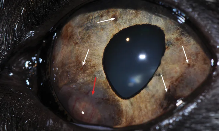
Nodular melanoma in the ventromedial aspect of the iris in a cat. The periphery of the mass is heavily pigmented (red arrow), whereas the center is poorly pigmented (asterisk). This melanoma invaded the ciliary body and the iridocorneal angle and caused secondary glaucoma. Multifocal iris pigmentation (white arrows) and dyscoria can also be noted.
Although uncommon, intraocular tumors can occur in dogs and cats, with primary anterior uveal melanocytic tumors being the most common.1-3 Melanocytic tumors may be malignant (ie, melanoma) or benign (ie, melanocytoma). In dogs, melanocytic tumors are twice as common as other primary intraocular tumors,2 and iris melanocytomas are approximately 3 times more common than melanomas.4,5
Clinical Signs
In dogs, iris melanocytic tumors arise from the anterior surface of the iris and are usually discrete, raised pigmented masses. As compared with ciliary body tumors, iris melanocytic tumors are easier to detect because of their anatomic location, thus allowing for earlier diagnosis in the course of the disease.2 In cats, flat, diffuse iris melanoma is more common than the nodular form (Figure 1) and usually starts as focal or multifocal areas of iris pigmentation, which may progress over time. Flat, diffuse iris melanoma cannot be differentiated from benign, diffuse iris melanosis based on clinical appearance alone (Figure 2; see Iris Freckles, Nevi, & Melanosis).6 Observation of clinical signs (eg, dyscoria, iris thickening, pigment dispersion, increased intraocular pressure) may help with the decision to enucleate an eye. In cats, some histologic and immunohistochemical parameters have been found to be helpful in determining the risk for metastasis of diffuse iris melanoma.7 Although these parameters may not be a practical tool for deciding whether to enucleate an eye, they may be used postenucleation to determine the metastatic risk in the individual cat and, when applicable, to form a tailored postoperative follow-up plan. Metastatic melanocytic tumors affecting the anterior uvea are rare but have been reported in dogs.8
For additional considerations regarding potential malignancy of diffuse iris pigmentary changes, see Iris Freckles, Nevi, & Melanosis.

Diffuse iris pigmentation progressing toward the iridocorneal angle in a cat. Diffuse iris melanoma cannot be differentiated from benign iris melanosis based on clinical appearance alone.
Predisposition
Breeds predisposed to melanocytic tumors include crossbreed dogs, Labrador retrievers, German shepherd dogs, golden retrievers, schnauzers, and cocker spaniels.5 Spayed dogs are also more likely to develop both benign and malignant melanocytic tumors.5 Middle-age to older dogs are more commonly affected.5 The median age of dogs at the time of ocular melanocytoma or melanoma diagnosis is 9 years.5 Inherited melanoma affecting young adult (ie, mostly between 1-2 years) Labrador retrievers has also been reported.2 No predispositions in cats have been identified.


A cluster of small, heavily pigmented uveal cysts over the iris and close to the pupil margins (A; white arrow) and free-floating in front of the pupil (yellow arrow) in a dog. Two big, heavily pigmented uveal cysts posterior to the pupil in a cat (B; arrows).
Diagnosis
Diagnosis of melanocytic tumors is based on typical appearance and clinical signs. Changes in iris color and/or a visible mass raised from the surface of the iris are typical. Additional clinical signs may include dyscoria as the mass progresses in size, hyphema, and, in advanced stages, uveitis due to tumor necrosis. Glaucoma may develop secondary to uveitis and/or due to invasion of the tumor into the iridocorneal angle and mechanical blockage of the aqueous humor outflow. Retinal detachment and blindness may also be noted in advanced stages.9
Darkly pigmented iridociliary cysts (Figure 3) may be confused with melanocytic neoplasia, particularly in cats.10 Ocular ultrasonography can be helpful in differentiating darkly pigmented cysts from uveal masses and should be performed prior to enucleation, especially in cats, as cysts are benign and do not require enucleation.


This aggressive iris melanoma in a dog obliterated most of the intraocular space, and the eye was enucleated (A). An aggressive amelanotic melanoma in a different dog (B) is present on the medial aspect of the anterior chamber, obliterating approximately one-third of the anterior chamber and causing dyscoria. Enucleation is indicated.
Treatment & Prognosis
The metastatic rate of anterior uveal melanoma is less than 5% in dogs.2 Thus, early intervention by means of referral to an ophthalmologist and referral procedures (eg, sector iridectomy, laser therapy11) should be considered in patients with focal lesions, with the aim of retaining the eye and vision. However, enucleation is advised in cases of fast-growing, locally invasive melanoma (Figure 4). Conversely, the metastatic rate of anterior uveal melanoma in cats is high (up to ≈60%),12 and early enucleation may prevent premature death.13 However, because diffuse iris melanoma cannot be clinically differentiated from benign melanosis, the decision to enucleate the eye can be challenging (see Iris Freckles, Nevi, & Melanosis).
Laser treatment of suspected diffuse iris melanoma in cats has been suggested to effectively delay tumor progression14 but remains controversial due to the difficulty in differentiating benign diffuse iris melanosis from melanoma.