Anesthesia in an English Bulldog
Jennifer E. Carter, DVM, DACVAA, CVPP, University of Melbourne
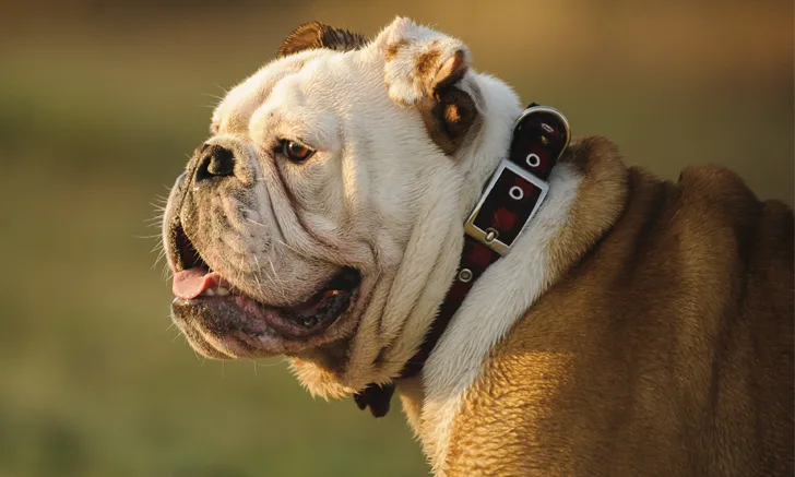
Hugo, a 7-year-old neutered male English bulldog, is presented for dental cleaning with possible extraction because of a suspected mandibular tooth root abscess. Hugo had been eating and drinking normally until 5 days ago, when his owner noticed he was less interested in his food and was drooling excessively.
At one year of age, Hugo was neutered and underwent soft palate resection, sacculectomy, and alarplasty of the nares. He has a 7-year history of snoring. He is up-to-date on core vaccinations. His 80-year-old owner does not leash walk him, and Hugo has access to a fenced yard for short times only for urination and defecation.
On physical examination, BCS is 7/9. Hugo is panting loudly and is excited but friendly. Although his excitement makes auscultation of the heart and lungs difficult, no obvious murmurs are evident. No other abnormalities are noted. Preoperative CBC results demonstrate a moderate neutrophilia (14.5 × 10³/uL [14.5 x 10⁹/L]; range, 3-12 × 103/uL [3-12 x 109/L]) with no left shift. Serum chemistry profile results are unremarkable.