Acute Respiratory Distress in a Brachycephalic Dog
Gretchen Statz, DVM, DACVECC, Antech Diagnostics, Veterinary Emergency and Specialty Care, Indianapolis, Indiana
THE CASE
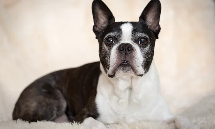
Yoyo, a 10-year-old neutered male Boston terrier, is presented to the emergency room for increased respiratory effort of 24 hours’ duration. On presentation, the dog starts coughing and gagging, then develops wheezing that progressively worsens. He had been eating normally before onset of respiratory distress.
Yoyo has always been overweight but otherwise healthy and is not on any medications. The results of a routine physical examination and laboratory work performed 6 months prior are unavailable but were reportedly normal according to the owner. Yoyo has no history of vomiting or regurgitation.
On physical examination, Yoyo is bright and alert. Temperature (101.2°F [38.4°C]) and heart rate (120 bpm) are within normal limits. There is mild serous discharge from the left eye and increased respiratory effort with loud respiratory stridor and soft stertor on inspiration. Respiratory rate is elevated (66 breaths/min), and Yoyo wheezes on exhalation. Heart sounds are muffled bilaterally. His abdomen is pendulous, and BCS is 7/9.
The owner agrees to CBC, serum chemistry profile, and thoracic radiography. CBC and serum chemistry profile are unremarkable aside from elevated liver values (Table). Airway sampling for cytology and culture was discussed but not pursued, likely because of the patient’s critical condition.
Table: Elevated Liver Values
Thoracic radiographs (Figure) show an alveolar lung pattern in the ventral aspect of the right middle lung lobe that is most consistent with aspiration pneumonia or bronchopneumonia. Hemorrhage, neoplasia, and bronchial obstruction are also on the differential list but are considered less likely. Hepatomegaly is present, as is radiographic evidence of possible palate/pharyngeal region swelling and aerophagia.

FIGURE Right lateral (A), left lateral (B), and ventrodorsal (C) thoracic radiographs of the patient. Palate/pharyngeal region swelling can be observed (B; arrow), as can an alveolar lung pattern in the ventral aspect of the right middle lung lobe (C; arrow), which is most consistent with aspiration pneumonia or bronchopneumonia.
Yoyo is admitted to the hospital, placed in an oxygen cage at a fraction of inspired oxygen (FiO2) content of 40%, and treated with ampicillin–sulbactam (22 mg/kg IV q8h), enrofloxacin (10 mg/kg IV q24h), terbutaline (0.01 mg/kg IV q12h1), and nebulization with 0.9% saline. He is placed on IV fluids at 50 mL/hr based on maintenance plus estimated subclinical dehydration, and his respiratory rate and effort are monitored every 2 hours.
What Are the Next Steps?
Disclosure: Dr. Statz is also affiliated with ANTECH Diagnostics.