Acute Onset of Alopecia & Erythematous Dermatitis in a Vizsla
A 4-year-old, neutered male Visla presented with a history of intermittent prur itus several years in duration and episodes of otitis externa.
History
Recently, large areas of alopecia, scaling, and erythema primarily involving the head and dorsal thorax were noted. The owners also reported that periocular, facial, and pedal pruritus were present. Prior treatments included a 3-week course of cephalexin (25 mg/kg Q 12 H), hydroxyzine (2.5 mg/kg Q 12 H) and prednisone (0.5 mg/kg), and a 10-week strict elimination diet trial with a duck and potato, restricted-antigen diet. During the trial period, no other foods or flavored medications were administered. The pruritus persisted, although it was greatly reduced with prednisone therapy. There was no improvement in the alopecia.

Physical Examination
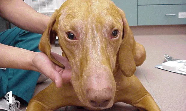
Physical findings were normal except for the ears and skin. Symmetrical alopecia was present on the face, dorsal head, dorsal neck, and thorax. Close examination of the skin revealed scaling and erythema (Figures 1 and 2). Hairs from affected areas were epilated easily, and a whitish-yellow debris was adherent to the proximal hair shaft/bulb (follicular casting). Bilateral otitis externa was present, characterized by thick, dark-brown ceruminous debris that obstructed both ear canals. Mild interdigital erythema and salivary staining were present on the feet.
ASK YOURSELF ...
On the basis of the clinical presentation, what are the most likely differential diagnoses?
What diagnostic tests do you recommend?
Based on this pet's diagnosis of both sebaceous adenitis and presumptive atopic dermatitis, what treatments do you recommend?
Diagnosis: Sebaceous adenitis
This patient had two major dermatologic problems: recurrent pruritus and alopecia. The history suggested that the problems were distinct and unrelated. Follicular casting, which is seen in many disorders of keratinization, and easily epilated hairs suggested that there was a disease process affecting the hair follicles. Common disorders affecting hair follicles include but are not limited to demodicosis, dermatophytosis, and staphylococcal pyoderma.
Staphylococcal pyoderma is uncommon on the face, but may be present in association with other disorders, such as demodicosis. Follicular casting is common in disorders of keratinization and sebaceous adenitis.
Differential Diagnoses
Differential diagnoses for the pruritus included infections (bacterial pyoderma, dermatophytosis, and Malassezia dermatitis), parasites (e.g., demodicosis, scabies), and allergies (atopy, food allergy, flea allergy), although failure to improve with a restricted, reduced-antigen diet reduced the possibility of a dietary sensitivity. The distribution of the pruritus was most compatible with an allergic cause or a parasitic dermatitis (scabies). Otitis externa is common with allergic dermatitis and is often complicated by infection with bacteria and/or yeast. Otitis externa may be seen secondary to other disorders (e.g., parasitism, endocrinopathy, etc.); however, based on the working diagnosis, we assumed the otitis in this patient was secondary to allergic disease.
Diagnostic Tests
Several deep and superficial skin scrapings from the head and feet were collected and evaluated microscopically for the presence of mites. No parasites were found. Dermatophytosis was ruled out based on a negative Wood's lamp examination, lack of finding ectothrix spores on a trichogram, and a negative fungal culture. The trichogram did reveal follicular casting at the base of the hairs. Three 8-mm punch biopsies were collected from affected areas, placed in formalin, and sent to a dermatopathologist for evaluation. Cytology samples were collected from the ears and evaluated microscopically. On a subjective grading scale of 0 to 4, 3+ yeast was found in both ears. Cytologic samples were collected from the skin using acetate tape. These samples were stained and evaluated microscopically. No organisms were found.
Negative findings on skin scrapings ruled out demodicosis. However, negative skin scrapings do not rule out scabies and, because of the distribution pattern, scabies was still considered a possible differential diagnosis, although allergies were considered more likely. The lack of a response to a food trial made food allergy unlikely. Facial and pedal pruritus are typical of atopic dermatitis and were considered the most likely cause of the pruritus. Atopy is a diagnosis based on clinical signs and, most important, ruling out other pruritic skin disorders. It is a diagnosis of exclusion. Atopy would also account for the yeast otitis. In-house diagnostic testing (skin scrapings and fungal infection investigations) did not reveal a cause of the hair loss. Because of the high suspicion of sebaceous adenitis, skin biopsy samples were collected to investigate the cause of the hair loss.

Treatment
Pending the skin biopsy findings, treatment included ear cleaning, topical otic therapy containing a steroid (fluocinolone), and dimethyl sulfoxide and whole-body treatment with a miconazole solution. Selamectin was also administered in the event of scabies.
Biopsy results revealed a multifocal granulomatous and pyogranulomatous dermatitis obscuring sebaceous glands. Lesions were consistent with granulomatous sebaceous adenitis. Sebaceous adenitis is a condition whose pathogenesis is not well understood. An inflammatory process leads to destruction of the sebaceous glands and alopecia ensues due to peripheral involvement. There is undoubtedly a genetic predisposition, as certain breeds tend to be affected-the disease is common in Vislas.
There is no cure for sebaceous adenitis. Some treatments have been found to be beneficial in some but not all affected animals. Treatment options include retinoid or vitamin A therapy, microemulsion cyclosporine, and topical therapy with benzoyl peroxide shampoos and propylene glycol rinses.
Treatment for this pet was selected after discussing options with the owner. Due to the presence of concurrent allergic dermatitis and sebaceous adenitis, cyclosporine therapy was selected. Treatment was initiated with 5.2 mg/kg cyclosporine PO once daily. The pet was reevaluated after 4 weeks of therapy. The owner reported a dramatic decrease in pruritus and discomfort. Erythema and scaling was reduced in the alopecic areas, but hair growth was not evident. Cyclosporine was continued at the same daily dose for an additional month. At that time, new hair growth was evident in all affected areas. The dosing was decreased to alternate-day administration and continued successfully for 3 months, at which time the dose was decreased to 3 times weekly. The pet continued to do well. Attempts to decrease the dosing to twice weekly have been unsuccessful. The pet continues to receive cyclosporine 3 times weekly after 2 years (Figure 3).
Did You Answer ...
Deep and superficial skin scrapings (mites), Wood's lamp examination, trichogram, fungal culture, punch biopsy, and otic and skin cytology samples
Initially: Ear cleaning, topical steroid, dimethyl sulfoxide, whole-body miconazole, and scabicide Sebaceous adenitis/pruritus: Options include retinoid or vitamin A, cyclosporine, benzoyl peroxide shampoo, and/or propylene glycol rinse