In the Literature
Seger CB, Regier P, Ham K, et al. Presentation, diagnosis, and management of gossypibomas in veterinary specialty hospitals: a multi-institutional study of 21 cases. Vet Surg. 2026;55(1):101-109. doi:10.1111/vsu.14307
The Research …
Gossypibomas are lesions that occur secondary to granulomatous inflammation from a textile (eg, surgical gauze, sponge) retained inside a patient’s tissue following surgery. Although rarely reported in veterinary patients, multiple case reports and reviews exist in human literature.1,2 Reviews of human medicine cases have shown that awareness of gossypibomas, use of systematic perioperative checklists, and use of radiopaque products are effective for preventing retained surgical items.3
This retrospective study reported the incidence, management, and outcome of gossypibomas in small animals. Gossypibomas were identified in the medical records of 18 dogs and 3 cats (n = 21). Ovariohysterectomy was the most common initial surgery leading to gossypiboma, and 95% of retained sponges were confirmed as located in the abdomen.
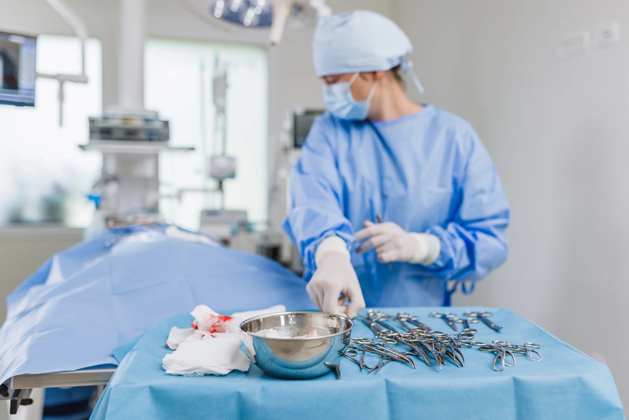
Pre- and postoperative sponge counts were performed in 19% (4/21) of cases, not performed in 19% (4/21) of cases, and not reported in 62% (13/21) of cases. Sponges containing radiographic indicators were used in 19% (4/21) of cases, not used in 38% (8/21) of cases, and not reported in 43% (9/21) of cases. Postoperative radiography was performed in 2 cases based on suspicion of sponge retention. In both cases, the sponge was identified radiographically and removed during the same anesthetic period. Median time from initial surgery to presentation was 13.5 days, with most patients presented within 30 days. Presenting signs (eg, pyrexia, lethargy, vomiting, anorexia) were nonspecific.
Abdominal ultrasonography and CT demonstrated elevated sensitivity for gossypiboma detection compared with other diagnostic modalities, usually displaying a mixed echogenic/attenuating mass; this elevated sensitivity has also been shown in prior research.4 Perioperative checklists were not reported in 80% (17/21) of cases, and gauze without radiographic markers was used in 67% (8/12) of cases, with gauze type recorded at initial surgery. Most patients had adhesions, and removal involved resecting multiple tissues (eg, gallbladder, kidney, small intestine) in some patients. Two patients did not survive to discharge.
The study authors concluded that surgical removal of gossypibomas can lead to good outcomes but may be associated with severe complications or death.
… The Takeaways
Key pearls to put into practice:
Gossypibomas should be considered in patients presented with nonspecific clinical signs and evidence of systemic and/or local inflammation, especially in patients that have recently undergone surgery.
Gossypibomas are preventable, and measures should be implemented for prevention, including a perioperative surgical safety checklist with a sponge count. Sponge count is not infallible but should be performed before surgery and before wound closure to identify discrepancies, as is done in human medicine.5
Radiopaque sponges are recommended for easier detection postoperatively. Prompt radiographic evaluation should be pursued if there is concern for a retained surgical object.
The American College of Surgeons recommends thorough wound evaluation before closure to prevent retained objects and help catch sponge counting errors that could offer a false sense of security.6 These practices can be adopted in veterinary medicine to prevent formation and improve detection of gossypibomas.
You are reading 2-Minute Takeaways, a research summary resource presented by Clinician’s Brief. Clinician’s Brief does not conduct primary research.