Ultrasonography in Practice: Practical Considerations
The use of ultrasonography in veterinary medicine has had a great impact in diagnostic accuracy and quality of care.
Over the past 10 years, technical advancements have increased the utility of ultrasonography and decreased its cost to general practitioners.<sup1–3sup> Before purchasing an ultrasonography unit, however, you should ask several important questions about anticipated application and expense:
• What types of ultrasonographic examinations will you be performing most frequently?• Will you mainly be doing abdominalultrasonography?• Is a member of your practice experiencedin echocardiography?• Will you be investigating lamenesses with musculoskeletal ultrasonography?
The answers to these initial questions will dictate how much you spend on your unit and which transducers you select for purchase.
It is important to note that the utility of ultrasonography is almost entirely operator dependent; the purchase of a technologically superior unit is no substitute for time, patience, experience, and expertise. Therefore, acquisition of any ultrasonography equipment should be accompanied by a regimented training program; review of related texts and articles;2,4 an understanding of cross-sectional anatomy; and frequent, practical application (practice, practice, practice) in your hospital.
IndicationsUltrasonography has many applications in veterinary medicine. Perhaps the most common is the abdominal examination, closely followed by echocardiography. The utility of ultrasonography in the diagnosis and management of musculoskeletal disease has also been well documented. Cervical ultrasonography, especially for evaluating the thyroid lobes, is another common application. Ocular ultrasonography can help evaluate the fundus in patients with mature cataracts. Noncardiac thoracic ultrasonography is also useful for assessing pleural disease, mediastinal masses, and peripheral pulmonary lesions (Figure 1).

(Figure 1: A radiologist performs a noncardiac thoracic ultrasonographic examination. Note that ambient lighting was increased for photography purposes; imaging should be performed in a darkened environment.)
One of the most helpful applications of ultrasonography in veterinary medicine is ultrasonography-guided fine-needle aspiration and biopsy. Often, the results of ultrasonography are nonspecific. The identification of parenchymal nodules or masses may indicate a benign or malignant process, and cellular evaluation is needed for definitive diagnosis. Ultrasonography is an excellent tool for safely and accurately guiding a needle into a nodule or mass to ensure proper sampling and an accurate diagnosis.
In addition, the use of ultrasonography to guide abdominocentesis, thoracocentesis, pericardiocentesis, cystocentesis, or biliary centesis increases the safety of each procedure when small volumes of fluid are sampled. In some cases of large fluid accumulations, ultrasonography may also produce a therapeutic effect.
Considerations
Complementary RoleUltrasonography is often incorrectly considered a superior imaging modality due to its ability to reveal the internal structure of an organ. Ultrasonography does allow for identifying distinct internal features of soft tissue structures and organs that are not possible to distinguish on radiography. For example, a radiographically enlarged kidney may indicate the presence of a renal mass, diffuse parenchymal enlargement, or hydronephrosis. Distinction between these soft tissue radiographic lesions may be possible with ultrasonography. However, the complementary role of diagnostic-quality abdominal radiography in the complete evaluation of suspected abdominal disease cannot be overstated. Diagnostic-quality radiographs supply information about the musculoskeletal system and help focus the ultrasonographic examination.
Operator SkillThe accuracy and utility of ultrasonography are totally operator dependent. It is the role of most veterinary ultrasonographers to produce diagnostic-quality images while simultaneously interpreting findings. Recognition of lesions that require further interrogation and the proper application of additional tools, such as color Doppler, are important in the thorough evaluation of the veterinary patient.
To accurately interpret ultrasonographic images, a working knowledge of ultrasonography physics, including ultrasound wave production, interaction of ultrasound waves with tissues, and ultrasonographic artifacts is necessary.1 Although many weekend short courses in ultrasonography instruction are available, these are brief introductions to ultrasonographic examinations and interpretation. Using a high-quality atlas4 and an anatomy text will help the successful ultrasonographer build skill in the examination. A focus on the production of diagnostic-quality images is essential. The wide availability of teleradiology consultation can aid in image interpretation.
Evaluating EquipmentUltrasonographic equipment is best evaluated by performing an ultrasonographic examination and directly comparing the images created by using the same imaging parameters (eg, depth, frequency). Image quality is primarily dictated by transducer quality and can be measured in terms of spatial resolution. Spatial resolution is the ability of the unit to distinguish 2 objects that are in close proximity as separate structures. This can be measured in the lateral (left-to-right on the image plane), axial (top-to-bottom on the image plane), and elevational (resolution in the plane of slice thickness) directions. The construction of a phantom may aid in comparing multiple ultrasonography units; inexpensive methods for building one have been described.5 Alternatively, examining the same patient with each ultrasonography unit may provide a standard for comparison.
Economic ImpactAlthough the cost of ultrasonography technology has decreased over time, the investment required for a high-quality ultrasonography unit is still significant. The practitioner must realistically assess the frequency with which the ultrasonography unit will be used and justify the expense. Experience will increase diagnostic accuracy and confidence, but it is the unfortunate habit of most veterinarians to neglect to charge for tests in which their confidence is lacking.
The Inner Workings of Ultrasound
The essential components of the ultrasonography unit are the transducer (Figure 2) and computer (Figure 3).

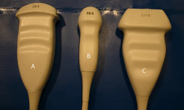
(Figure 2: Ultrasound transducers--convex broadband transducer that can transmit at a frequency range of 9-4MGz (A); microconvex transducer with a small footprint that can transmit at a frequency range of 8-5 MHz (B); linear transducer that can transmit at a frequency range of 17-5 MHz (C).)
At the heart of the transducer is the piezoelectric crystal, a substance that reacts to the periodic application of an electrical charge to produce the ultrasound waves used for imaging. The frequency of ultrasound waves produced by the crystals, as well as the configuration of these crystals, dictates the application of each transducer. Frequency is directly proportional to spatial resolution and is inversely proportional to penetration:
• High-frequency transducers are typically used in smaller patients or to visualize superficial structures (tendons, thyroid lobes, feline abdomens)• Low-frequency transducers are used if spatial resolution can be sacrificed for penetration and increased imaging depth (abdominal examinations in large dogs, thoracic examinations in horses). Common crystal configurations, or crystal arrays, include linear-, sector- (phased-), convex-, and microconvex-array transducers:• Linear transducers consist of a straight, flat array of crystals that produce a rectangular field of view, with a wide, flat footprint. They are typically high-frequency transducers, providing high spatial resolution but more limited penetration.• Sector (phased-array) transducers consist of an array of crystals that rapidly produce ultrasound waves and thus yield images with high temporal resolution; a small, square footprint; a pie-shaped field of view; and some loss of spatial resolution. These transducers are most commonly used in echocardiography but can be used in other areas as well.• Convex and microconvex transducers offer a compromise between the spatial resolution of a linear configuration and the small footprint and wide, pie-shaped field of view provided by a sector transducer. Convex transducers are slightly larger than microconvex transducers, and may be more difficult to maneuver when scanning a patient. Microconvex transducers offer a smaller footprint, and often allow for easier and more complete evaluation of the cranial abdomen.

(Figure 3: Ultrasound unit with a flat-screen display, 4 transducers--2 linear array, 1 convex, and 1 microconvex--a touch screen, and a relatively simple ergonomic design. A keyboard is located beneath the interface to allow for additional image labeling.)
The type or types of transducers purchased will be directly influenced by the applications most frequently performed. In all cases, a compromise is made between spatial resolution and penetration, and the sonographer must select the proper transducer for each application. For ultrasonographic evaluation of the abdomen, a microconvex high-frequency probe, a convex lower-frequency probe, and a linear high-frequency probe would be an ideal place to start. This would allow the practitioner the greatest flexibility for the various abdominal applications based on patient size and shape. For practices limited to cats or small dogs, the lower-frequency convex transducer would not be necessary.

ULTRASONOGRAPHY IN PRACTICE: PRACTICAL CONSIDERATIONS • Matthew D. Winter
References1. AAPM/RSNA physics tutorial for residents—B-mode US: Basic concepts and new technology. Hangiandreou N. Radiographics 23:1019-1033, 2003.2. Small Animal Diagnostic Ultrasound. 2nd ed. Nyland T, Mattoon J—Philadelphia: WB Saunders, 2002.3. Practical and financial considerations that affect selection and purchase of ultrasound equipment. Roberts W. Obstet Gynecol Clin North Am 25:663-676, 1998.4. Atlas of Small Animal Ultrasonography. Penninck D, d’Anjou M—Ames, IA: Blackwell Publishing Professional, 2008.5. Anatomic models and phantoms for diagnostic ultrasound instruction. Carrig C, Pyle R. Vet Radiol Ultrasound 42:320-328, 2001.