Selecting the Right Blood Collection Tube for Diagnostic Testing
Rachel Victoria Poulin, RVT, VTS (SAIM), Wheat Ridge Animal Hospital, Wheat Ridge, Colorado
Veterinary nurses are responsible for obtaining and submitting patient blood samples for diagnostic testing, and they must be familiar with laboratory test requirements to ensure that correct sample types and amounts are collected in appropriate tubes. Improper collection and tube selection can adversely affect test results, causing potential misdiagnosis and delaying patient treatment.
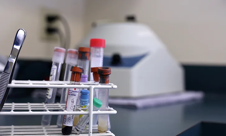
Blood collection tubes are distinguished by top color, which identifies tube additives. Tube additives prevent or activate clotting and determine the final blood product (eg, whole blood, serum, plasma) submitted for testing. Anticoagulant tubes include lavender (purple), green, and light blue. Tubes with red and red/gray (marble or tiger) tops do not contain anticoagulant additives. (See Table 1.)
Blood Collection
Select a syringe that can hold the amount of blood needed for the test, using a 22-gauge or larger needle depending on the patient’s size.1 If a smaller needle must be used, aspirate the blood from the vein slowly to avoid hemolysis.1 Perform venipuncture as quickly and smoothly as possible to prevent rupture of red blood cells2 and minimize patient stress. If multiple attempts are necessary, use a new needle each time.
Once the correct amount of blood has been obtained, remove the needle from the syringe and the top from the tube, and slowly eject the syringe contents into the tube using gentle, even pressure. If vacuum tubes are used, insert the needle, with the syringe attached, through the rubber stopper and allow the negative pressure in the tube to pull the blood from the syringe.3 If multiple vials of blood are required, ask a team member to help manipulate the tubes correctly to reduce the need for multiple venipuncture to the patient. A vacuum collection system may also be used to fill tubes directly.
Fill each tube with the amount of blood required for the test. If too little blood is added to a tube with an anticoagulant, cell damage may occur because of anticoagulant overconcentration.1,3 The amount of anticoagulant in vacuum tubes is based on tube size,1 so allow these tubes to fill automatically, ensuring an adequate amount of blood for the size of the tube. When obtaining blood for multiple tests, fill the tubes in the following order: light blue, red, red/gray (marble or tiger), green, and lavender (purple).4
Blood Collection Tubes for Diagnostic Testing
, Almost all tubes need at least 8 to 10 inversions or they need to be on a rocker.12
Gently invert tubes that contain an anticoagulant to ensure mixing of the anticoagulant and blood; follow suggested inversion times for each tube color. (See Table 1.) Using an automatic tube rocker is useful for ensuring gentle, even mixing. If an automatic tube rocker is not available, gently rocking a sample that has been sitting is imperative. Do not freeze whole blood, unseparated serum, or unseparated plasma.2 Clearly label tubes with the patient’s name, signalment, and date of collection.
Whole Blood
Whole (unclotted) blood consists of plasma, red and white blood cells, and platelets and is required for complete blood counts, blood typing, and blood film examination.2 Whole blood is collected in lavender (purple) and green top tubes and is not centrifuged before submission to the diagnostic laboratory.
Lavender (Purple) Top Tubes
Lavender (purple) top tubes contain EDTA, an anticoagulant that works by chelating calcium, which is necessary for clot formation.5 Because EDTA does not alter cell morphology, it is the preferred anticoagulant for hematologic studies in dogs and cats.6,7 Make sure the blood sample is mixed well with the EDTA to avoid sample clotting. Do not shake the tube, as this can damage cell structure and integrity.6 Use lavender (purple) top tubes to submit whole blood for tests that examine red and white blood cells and platelets.
Green Top Tubes
Green top tubes contain heparin, an anticoagulant that acts by inhibiting thrombin formation.8
Serum
Serum samples are required for blood chemistry panels, serology, immunology, and most endocrinology tests.2 Serum is the fluid portion of the blood that has had fibrinogen removed during clotting. Blood for serum testing is collected in red or red/gray (tiger or marble) top tubes.
Red & Red/Gray (Tiger or Marble) Top Tubes
These are serum separator tubes (SST) and are commonly used for any test requiring serum, with the exception of drug monitoring testing.9 Plastic red top and marble (tiger) top tubes contain a gel to separate serum from the clot and a clot activator but no anticoagulants or preservatives. Let the sample sit in a vertical position for 15 to 20 minutes at room temperature, then centrifuge at 2500 revolutions per minute for 10 to 15 minutes.9
Plasma
Plasma is required for blood chemistry, coagulation, platelet function, and some toxicology tests.2 Plasma is the liquid portion of the blood from which the red and white blood cells and platelets have been removed. Plasma consists primarily of water with dissolved proteins, hormones, lipids, enzymes, salts, carbohydrates, vitamins, and waste materials.10 Lavender (purple), blue, and green top tubes are used to collect samples for plasma testing. Once the appropriate amount of blood has been placed in the tube, centrifuge it as soon as possible for 10 minutes9 and transfer the supernatant to a clean, labeled tube with no additives for submission to the diagnostic laboratory.
Green Top Tubes
Green top tubes contain heparin; once centrifuged, the supernatant is considered heparinized plasma. This tube should be used for blood chemistry analysis and toxicology testing for some metals (eg, copper, iron, zinc) and nitrates and nitrites.2
Light Blue Top Tubes
Light blue top tubes contain an anticoagulant called sodium citrate, which removes calcium from the blood to prevent clotting. If serum is necessary for a requested test, place the sample in the tube and mix gently; centrifuge as soon as possible, and transfer the supernatant into the appropriate testing tube.2 Sodium citrate tubes are used for most comparative coagulation testing because the anticoagulant is reversible. Calcium can be added back into the sample to study coagulation in a controlled environment.11 Use light blue top tubes for testing prothrombin time, activated partial thromboplastin time, and fibrinogen.3
Conclusion
Blood samples must be properly collected, handled, and labeled to ensure accurate diagnosis and treatment of veterinary patients. Veterinary nurses must be familiar with the sample requirements (including proper tube, sample amount, and sample type) of each diagnostic test ordered by the veterinarian.