In the Literature
Charlesworth T, Sampaio E. Effect of hospitalisation on the rate of surgical site infection in dogs with Penrose drains. J Small Anim Pract. 2023. doi:10.1111/jsap.13678
The Research …
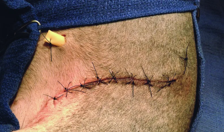
Passive drainage systems (eg, latex Penrose drains) are inexpensive and can be used to drain wounds and reduce seroma formation. Penrose drains function via gravity and capillary action, with fluid clinging to the exterior surface of the drain and exiting through a ventral opening in the skin that may cause risk for ascending infection.1-3
This retrospective study reviewed complication and infection rates in dogs (n = 208) that underwent Penrose drain placement in wounds following a variety of surgeries, including traumatic wound repair, abscess drainage, mass removal, and sialoadenectomy. Most drains were placed with the dorsal end held subcutaneously in the deepest, most dorsal portion of the wound by a subcutaneous absorbable or external transcutaneous suture and the ventral end exited through a fresh incision and secured to the skin with a suture.
At least 53.4% of dogs were fitted with an Elizabethan collar and/or protective garment, and at least 62.5% had an absorbable dressing applied. Postoperative antibiotics were administered to 88.5% of dogs. Drains were left in place for a median of 3 days, with 66.7% of dogs discharged within 24 hours of surgery with the drain in place. Postoperative complications were noted in 40.9% of dogs, with the most common being infections, seromas, dehiscence, and continued drainage. Complication rates were similar whether dogs were kept in the hospital or discharged.
… The Takeaways
Key pearls to put into practice:
The traditional drain placement technique was used in most dogs in this study. A dorsal external suture is helpful because it keeps the drain in the pocket and facilitates finding the drain if the ventral end retracts into the wound due to suture failure or being chewed off by the patient.1,2,4 A single drain exit point is typically recommended, except when limb movement (eg, in the axilla) results in air entrapment.1,2,4 The exit site should be ventral based on the patient’s standing position, separate from the wound closure to prevent incisional irritation, and larger than the drain in diameter to facilitate fluid flow.1,2,4
Covering Penrose drains with a sterile bandage is commonly recommended. In areas that are difficult to bandage, drains can be covered with absorptive gauze and an adhesive drape or a tie-over bandage.5 Bandages should be changed routinely to reduce risk for infection and monitor fluid production.1,2,4
Postoperative antibiotics are not routinely prescribed for patients with clean surgical wounds due to risk of selection for antimicrobial drug resistance.6,7 Drains are usually removed when the amount of drainage decreases and fluid becomes clearer.1,2,4 Risk for infection may increase if drains are left in place >3 days.3
At-home management of dogs with Penrose drains does not appear to increase complications and may be less stressful for patients, less expensive for pet owners, and safer for veterinary practices, particularly if a patient is aggressive or has a multidrug-resistant infection.7,8 Instructions should be provided to owners if patients are discharged with Penrose drains. The sterile covering should be changed daily, when it is loose, or when it has strike through (ie, visible fluid in the exterior layer). Owners should wash their hands, put on gloves, remove the bandage, clean the exit site with an antiseptic, and place a thin layer of triple antibiotic ointment on any irritated skin before placing a new bandage.4
You are reading 2-Minute Takeaways, a research summary resource presented by Clinician’s Brief. Clinician’s Brief does not conduct primary research.