Narrowed Palpebral Fissure in a Dog
Laura O'Sullivan, VMD, Media Veterinary Hospital, Media, Pennsylvania
Eric N. Glass, DVM, MS, DACVIM (Neurology), Red Bank Veterinary Hospital, Tinton Falls, New Jersey
Marc Kent, DVM, DACVIM (Internal Medicine, Neurology), University of Georgia
Alexander de Lahunta, DVM, PhD, DACVIM (Neurology), DACVP (Hon), Cornell University
Hansel, an 8-year-old neutered male German shepherd dog, was referred for persistent squinting and an inability to blink his right eye (OD).
History
Signs were present for 1 week. Hansel was otherwise normal, was current on vaccinations and heartworm and ectoparasite preventives, and had no pertinent medical history (eg, exposure to toxins, history of trauma). No blepharospasm was reported by the owner.
Examination
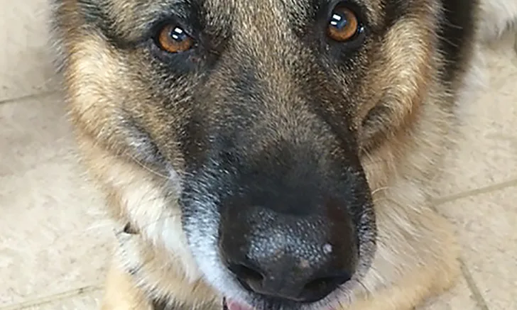
Figure 1. At presentation, the palpebral fissure is narrowed on the right (arrow) as compared with the left. The nasal planum is deviated to the right (arrowhead). The right ear is positioned slightly more caudal on the head as compared with the left ear.

Physical examination revealed deviation of the nasal planum to the right and slight elevation of the right ear (Figure 1). Otoscopic examination was normal. Neurologic examination included evaluation of mentation, gait, postural reactions, spinal reflexes, tone, and muscle mass in all limbs; all were within normal limits. Evaluation of the cranial nerves revealed that the palpebral reflex and menace response were absent OD. A narrowed palpebral fissure OD was noted. Response to stimulation of the nasal mucosa was normal. All other cranial nerve reflexes were normal.
Related Article: Step-by-Step: The Neurologic Examination
Diagnostics
CBC, serum chemistry panel, thoracic radiographs, and thyroid hormone testing (thyrotropin and free thyroxine by equilibrium dialysis) were normal. MRI of the head was normal.
Diagnostic Considerations
Anatomic diagnosis: Facial nucleus in the medulla or the facial nerve (cranial nerve VII) or hemifacial tetanus (HT) of the muscles of facial expression, including the obicularis oculi muscle (narrowed palpebral fissure) and levator nasolabialis (deviated nasal planum), or denervation atrophy (contracture) of the facial muscles. The lack of other neurologic deficits suggests the lesion did not involve the medulla.
Differential diagnoses: From the medulla, the facial nerve exits the cranial cavity via the internal acoustic meatus to enter the facial canal within the petrosal portion of the temporal bone. The facial nerve exits the facial canal via the stylomastoid foramen. Once extracranial, the facial nerve courses rostrally just ventral to the external ear canal where it branches. Causes to consider include disease of the middle ear, (eg, otitis media, which secondarily affects the facial nerve), or primary diseases of the facial nerve. Primary diseases include inflammation, neoplasia, aberrant blood vessel compressing the facial nerve, and trauma.
Diagnosis
Canine idiopathic HT
Related Article: Facial Nerve Paralysis
Discussion
Figure 2. On anesthetic induction for MRI, deviation of the nasal planum was not present (arrowhead). The dogs closed eyelids make it difficult to appreciate the narrowed palpebral fissure. Note the near-symmetrical positon of the ears.

During general anesthesia for the MRI, the patients narrowed palpebral fissure, deviation of the nasal planum, and abnormal ear position resolved (Figures 2 and 3). On recovery from anesthesia, the abnormalities returned. This confirmed HT1,2 and excludes from consideration denervation atrophy (contracture), as relaxation of the muscles under anesthesia would not occur.
Figure 3.A transverse plane, proton density MRI at the level of the medulla and rostral cerebellum of the patient. The normal facial and vestibulocochlear nerves are seen exiting the internal acoustic meatus (open arrows). Normal cochlea are seen bilaterally (solid arrows). The normal tympanic cavities are filled with air (asterisks).

HT results in persistent contraction of the facial muscles innervated by the facial nerve. A narrowed palpebral fissure, deviation of the nasal planum toward the affected side, and a caudal position of the ear are seen. Under general anesthesia or with local anesthesia of the facial nerve, muscle activity is inhibited, muscle relaxation ensues, and the patient appears normal.1 In some cases, ipsilateral, hemifacial paralysis, which then resolves, may precede HT. Used synonymously, hemifacial spasm is a misnomer because muscle spasms are not present.
In humans, HT is associated with microvascular compression of the facial nerve by aberrant vasculature. Treatment options (eg, botulinum toxin injections into the affected muscles, surgical microvascular decompression) exist.3 HT has been reported in 2 dogs secondary to a brain lesion.4 In the authors experience, HT in dogs mostly occurs secondary to an irritative lesion of the facial nerve. Irritative lesions likely cause excessive activity (depolarization) of the nerve rather than a lack of function.1
Diagnostics are aimed at identifying a metabolic disorder or structural lesion of the nerve or its nucleus in the medulla. An otoscopic examination should be performed to evaluate for abnormalities in the external ear canal because the facial nerve runs just ventral to it. A diagnostic evaluation consisting of CBC, serum chemistry panel, urinalysis, and thyroid hormone testing should help identify metabolic causes associated with nerve dysfunction. Finally, MRI of the head is used to evaluate the medulla and the course of the facial nerve. If an underlying cause is not identified, HT is assumed secondary to an irritative lesion. In such cases, supportive treatment aimed at muscle relaxation or empiric therapy for otitis media is not necessary.
Treatment
Given the normal results of the diagnostic testing and presumptive diagnosis, treatment was not pursued.4 Although the underlying cause is still not fully understood, it appears to remain a benign condition. Owners should be warned that the signs can remain indefinitely.
Related Article: Cerebral Infarction
CBC = complete blood count, HT = hemifacial tetanus, MRI = magnetic resonance imaging, OD = oculus dexter (ie, right eye)