Multiple Oral Pathologies in a Dog
Jerzy Pawel Gawor, DVM, PhD, DAVDC, DEVDC, Arka Veterinary Clinic Krakow, Poland
Cedric Tutt, BVSc(Hons), MMedVet(Med), DEVDC, MRCVS, Cape Animal Dentistry Service, Cape Town, South Africa
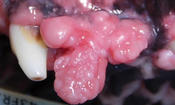
Gingival masses on the left rostral maxillary area covering buccal surface of premolar dentition
The Case
A 6-year-old, 55-lb (25-kg) spayed boxer was presented for a dental consultation regarding gingival masses (Figure 1) identified 2 months prior. Temperature, heart and respiratory rates, and the remainder of the physical examination were within normal limits. Clinical assessment and preanesthetic diagnostic investigation, including CBC and serum chemistry profile, were also within normal limits. During the examination of the patient’s head and oral cavity, a class III malocclusion was confirmed. Although this type of occlusion is typical for brachycephalic breeds, the maxillary incisors in this patient were causing trauma to the mandibular oral mucosa lingual to the mandibular incisors. The patient did not show signs of brachycephalic obstructive airway syndrome.
Diagnostics
A thorough evaluation of the oral cavity (Figure 2) was performed with the patient under general anesthesia. Mandibular first premolars were not visible on clinical inspection. Mandibular mucosal trauma by the maxillary incisors and trauma to the left mandibular canine (304) tooth by the left second maxillary incisor (202) were observed. The patient also had oral masses present at the gingiva of the left rostral maxillary area covering the buccal surface of the premolar teeth (Figure 1).
Full-mouth radiographs were obtained, with particular attention on the clinically missing mandibular premolars and the areas affected by gingival masses. Radiographs revealed unerupted mandibular 1st premolars situated in apparent cystic lesions, expansion of which resulted in involvement of the mandibular canines and 2nd premolar teeth. The apical portion of the mesial root of the mandibular premolar (306) was partially resorbed. Maxillary premolars were rotated bilaterally, but there were no periodontal consequences. Incidental findings included a supernumerary maxillary left first premolar 1 (205) and fusion of the roots of the left and right mandibular premolar 2 teeth (306 and 406). Horizontal alveolar bone loss was present at the area of 206, with radiolucency of the furcation area of 206 (Figures 3-6); the buccal aspect of 206 was covered by the gingival mass. Probing of the furcation was possible only from the palatal side and did not reveal furcation involvement.

FIGURE 2
Oral examination revealed nonvisible mandibular first premolars (stars), mandibular mucosal trauma by the maxillary incisors (arrows), and trauma to the left mandibular canine tooth (304) by the left second maxillary incisor (202; circle).
Treatment & Management
The following treatment plan was initiated based on clinical findings:
Extraction of unerupted mandibular premolars, debridement of cysts, and submission of removed tissue for histopathologic evaluation. Due to lack of definitive histopathologic diagnosis and no mobility of 306 and 406, the pet owner declined extraction of these teeth until planned follow-up.
Odontoplasty to reduce the crown height of the maxillary incisors to prevent trauma to the soft tissue and canine teeth. An alternative treatment option would include extraction of involved teeth. Radiographically, bone was present around the apices of 306 and 406; however, there was no indication to extract these teeth, as the cyst lining had been removed and thus new bone would fill the defect within a couple months.
Excisional biopsy of gingival enlargement at the area of the left maxillary canine tooth and submission for histopathologic assessment, followed by gingivoplasty to correct the size and shape of the gingival tissue.
The procedures were routine and without complications. The bony void where the cystic lesion was removed was allowed to fill with blood prior to the site being sutured closed, as this promotes primary healing, which includes the formation of new bone. The patient’s size and limited extent of bone damage did not require use of grafting material to fill the empty space after cyst removal. A CO2 laser was used to perform excisional biopsy of the gingival enlargement to decrease surgical time. After odontoplasty, there was no longer contact between the maxillary incisors and oral mucosa or the mandibular canine tooth. The maxillary incisor teeth that had undergone crown reduction were sealed with a dental varnish to protect the newly exposed dentin. An analgesic (ie, meloxicam [0.2 mg/kg IM, followed by daily oral doses of 0.1 mg/kg for 5 days]) was prescribed and application of an oral cleansing gel containing zinc ascorbate was advised for 14 days postoperatively.
Dentigerous cysts on the mandibular lesion and focal fibrous hyperplasia of the maxillary mass were confirmed via histopathology postoperation. At the 2-week postoperative recheck, the histopathology results were discussed with the owner and home care and further monitoring were established. Gingival enlargement may be plaque-associated or of a benign or malignant nature, and, because they are commonly found in boxer dogs, it is prudent for the owner to monitor the gingiva regularly. Plaque-associated gingival masses can be controlled with daily tooth brushing. Other benign or malignant lesions will commonly regrow following excision.
Discussion
Brachycephalic breeds are predisposed to certain pathologies, including dental retention, dentigerous cysts, and neoplastic and benign gingival enlargement.1-5 Anatomic features of brachycephalic breeds typically lead to painful traumatic malocclusion, which can only be confirmed via thorough oral examination. Because maxillary teeth crowding and gingival enlargement are strong causative factors for periodontal disease, this condition may develop sooner in brachycephalic breeds.
Systematic daily home care and regularly performed professional dental care can slow the return of gingival enlargement. It is essential to submit all excisions for histopathologic evaluation, as peripheral odontogentic fibroma, acanthomatous ameloblastoma, and gingival enlargement may have the same presentation.6
Approximately 30% of retained teeth result in odontogenic (ie, dentigerous) cysts, which destroy and weaken the bone, may cause pain, and can cause complications such as pathologic fractures or malignant transformation.5-7 Clinically missing teeth require radiographic evaluation, and intervention, if needed, should be prompt.7
Conclusion
Many pathologic situations, such as the one described in this article, are typically ignored and/or underestimated by owners and primary care clinicians. Despite appearing painful and bothersome, these pathologies may not cause significant behavior changes. Thus, thorough oral examinations should be performed regularly in all brachycephalic patients to assist with early diagnosis and prevention of periodontal disease.
Ask Yourself...
Multiple Oral Pathologies in a Dog Quiz
Take this quiz by answering the following multiple choice questions.