Laparoscopy Instrumentation
The term laparoscopy denotes that a procedure is performed completely under endoscopic guidance, negating the need for large incisions to enter the abdomen.
Although the advancement of minimally invasive surgery in veterinary medicine lags behind that of human medicine, descriptions of clinical techniques for laparoscopy, thoracoscopy, and arthroscopy in animals have increased dramatically in the past 5 to 10 years. This advancement has largely been driven by a changing client base that recognizes the benefits of less-invasive alternatives as well as clinicians who are motivated to learn new procedures and provide cutting-edge techniques to their clientele.
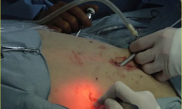
Figure 1 (View larger image): This dog is in dorsal recumbency and a pneumoperitoneum has been established. A is the camera portal placed midline just caudal to the umbilicus, B is a 5-mm instrument portal, C is the location of the second instrument portal, and * is the area of interest; A, B, and C are triangulate to maximize the accessibility of the area. Note the transillumination at C; the light source is used intracorporally to identify and avoid any superficial blood vessels during port placement.

Intestinal Endoscopic Biopsy Techniques
Basic PrinciplesLaparoscopy involves accessing the abdominal cavity through small incisions, creating a sufficient work space, and maneuvering within the abdomen using instruments activated extracorporally (Figure 1). Laparoscopic procedures currently performed in small animals include, but are not limited to, intestinal biopsies, intestinal foreign body removal, cystotomy, and gastropexy.

Instrumentation

The basic equipment needed to start minimally invasive surgery is listed in the Table. A basic start-up package typically costs $20,000 to $30,000; however, the equipment is the same as that used for thoracoscopy, so it may be possible to start laparoscopy and thoracoscopy without a large initial investment.
Figure 2 (View larger image): Standard tower setup for laparoscopy: A is the CO2 insufflator, intraabdominal pressure monitor, and CO2 flow regulator (L/min); B is the digital recording equipment, C is the monitor, and D is the halogen light source.

Figure 3 (View larger image): A) Typical laparoscopy instrument tray with both standard and laparoscopy instruments (A);
B) 5-mm right-angle endoscopic forceps being used for laparoscopic ureteronephrectomy__;

C) 5-mm fan retractor used for retraction of bowel or other viscera (the fan blades retract into a 5- or 10-mm sheath until deployed within the abdomen)

D) 5-mm endoscopic Metzenbaum scissors (top) and 5-mm endoscopic suture or hook scissors (bottom)__;

E) Veress needle, which is used for the blind or closed technique for establishing a pneumoperitoneum__;

F) 5-mm endoscopic spoon biopsy (top), which is used for liver and spleen biopsies, and a 5-mm curved hemostat (bottom), which can be used for blunt dissection and for grasping fine tissues
EndoscopesThe most commonly used endoscope is a 5-mm, 0-degree rigid endoscope. A 0-degree scope provides the surgeon with a visual field that is in line with the true field, makes orientation and manipulation of instruments easier, and facilitates light transmission better than those with an offset viewing angle. The most common angled endoscope is a 30-degree scope. This type enables visualization over the top of organs and widens the area of visualization.
At the end of the eyepiece, a camera unit is connected to a high-definition TV monitor. The camera can be sterilized using low-temperature hydrogen peroxide gas plasma technology (sterrad.com) or ethylene oxide, or it can be placed into a disposable sterile camera bag.
The endoscope must be attached to a high-quality light source. A high-intensity xenon light source is recommended because it gives the truest organ color, but less-expensive halogen light sources are also available.
CO2 InsufflatorA CO2 insufflator is used to create the pneumoperitoneum required to perform laparoscopy. Most systems have a standard means of CO2 delivery, an adjustable flow rate, and a constant or intermittent pressure reading. Intraabdominal pressure should be kept between 8 to 15 mm Hg. The author insufflates to the lowest pressure that allows the procedure to be done efficiently and safely. This is generally about 10 to 12 mm Hg.
Trocar-Cannula UnitsTrocar-cannula units are used to gain access to the body cavity, to provide a means to insufflate, and to maintain insufflation with a valve system and gas. The trocar is removed after the surgeon has accessed the cavity, and an endoscope or instrument is replaced into the remaining cannula. The cannula maintains access into the cavity and prevents unnecessary trauma to the body wall from repeated removal and insertion of instruments.
These units come with sharp or blunt trocars and correspond to the size of the instruments to be used (Figure 4). There are a variety of types of cannula subdivided into closed and open cannulae. Closed cannulae have internal valves or diaphragms that prevent leakage of gas when instruments are being switched out. Open cannulae allow leakage of the pneumoperitoneum and prolong the procedure time since it is necessary to occasionally reinsufflate; they are most commonly used for thoracoscopy since there is no need for insufflation.
Cannulae can also be rigid or soft. The rigid type is the most commonly used during laparoscopy and provides protection to the endoscope. Soft cannulae are generally used during thoracoscopy only to prevent trauma to the intercostal neurovascular bundle. Threaded cannulae are also available and can be helpful during thoracoscopic procedures because they tend to remain locked in place reliably.
Additional EquipmentA palpation probe is essential for moving and palpating abdominal and thoracic organs. Most probes have centimeter markings etched on them to allow the surgeon to measure the relative size of organs and masses.
For biopsies, oval biopsy cup forceps (liver, spleen, pleura, lymph nodes, masses), punch biopsy forceps (pancreas), and tru-cut style (core) biopsy needles (kidney, deeper tissues) are recommended. Endo-loops are valuable for biopsy of liver, tips of the pancreas, and spleen, and for some laparoscopic procedures, such as cryptorchidectomy.
Laparoscopic retrieval bags (ie, Endobag, covidien.com) can be useful for removal of large masses. These bags permit maceration of the tissue to reduce the size, thereby permitting removal through a portal site, or a minimally extended portal site, and thus can help prevent seeding of the body wall with potentially malignant cells.
Many instruments have a capability for monopolar electrocautery propagation near the handle. In addition, the harmonic scalpel and other vessel sealing devices (Ligasure, ligasure.com and EnSeal,surgrx.com) are available for minimally invasive surgical use.
Various grasping forceps, retractors, needle drivers, and scissors are also helpful.
Figure 4 (View larger image): Nondisposable trocar and cannula system for laparoscopy: 7-mm blunt trocar (top), 5-mm sharp trochar (middle), and 10-mm sharp trocar (bottom). Both blunt and sharp trocars are available for each cannula; having both types for each size cannula is recommended.

StaplersTypical clip applicators and stapling devices tend to be 10-mm or greater in diameter and require correspondingly larger trocar-cannula units. Thus, smaller stapling equipment designed specifically for minimally invasive surgery is available, as are several stapling devices for advanced procedures.
The endoscopic gastrointestinal anastomosis stapler simultaneously applies 6 rows of staples and simultaneously cuts between 3 rows of staples on the organ and 3 rows on the section to be removed. The stapling instrument has an articulating head that allows for correct placement of the staple line. The disposable staple loads come in 3 lengths—30, 45, and 60 mm—with staples sized 2.0, 2.5, 3.5, or 4.8 mm. The stapler itself can accommodate all cartridge sizes.
ConsiderationsThere are some inherent disadvantages to minimally invasive procedures. The equipment required to perform these procedures remains expensive. There are, however, many companies that market directly to veterinarians and refurbished systems are available for reduced cost. In addition, disposable instruments are always sharp and in excellent working order, but can be expensive; nondisposable products are less expensive, but can be costly to repair.
Many laparoscopic techniques are challenging to perform, and acquiring the necessary hand–eye coordination can be frustrating. Minimally invasive surgery involves different visual and tactile skills than traditional surgery; therefore, many veterinarians choose to refer rather than to obtain further training. For most advanced procedures, a large room and surgical team are necessary. Many procedures require a camera operator and 1 to 2 instrument operators.
Although being confident and competent with minimally invasive surgery requires a large time and financial commitment, it is rewarding professionally, financially, and personally.
LAPAROSCOPY INSTRUMENTATION • Stephen J. Mehler
Suggested Reading
A rapid and strong laparoscopic-assisted gastropexy in dogs. Rawlings CA, Foutz TL, Mahaffey MB, et al. Am J Vet Res 62:871-875, 2001.2. Cardiopulmonary effects of using carbon dioxide for laparoscopic surgery in dogs. Duke T, Steinacher SL, Remedios AM. Vet Surg 25:77-82, 1996.3. Comparison of laparoscopic ovariohysterectomy and ovariohysterectomy in dogs. Davidson EB, Moll HD, Payton ME. Vet Surg 33:62-69, 2004.4. Diagnostic quality of percutaneous kidney biopsy specimens obtained with laparoscopy versus ultrasound guidance in dogs. Rawlings CA, Diamond H, Howerth EW, et al. JAVMA 223:317-321, 2003.
Evaluation of laparoscopic-assisted placement of jejunostomy feeding tubes in dogs. Hewitt SA, Brisson BA, Sinclair MD, et al. JAVMA 225:65-71, 2004.
Laparoscopy. Monnet E, Twedt DC. Vet Clin North Am Small Anim Pract 33:1147-1168, 2003.7. Laparoscopy: Instrumentation and technique. Magne ML, Tams TR. In Tams TR (ed): Small Animal Endoscopy, 2nd ed—St. Louis: Mosby, 1999, pp 397-408.
Minimally invasive surgery: Laparoscopy and thoracoscopy in small animals. Remedios AM, Ferguson J. Compend Contin Educ Pract Vet 18:1191-1199, 1996.
Prospective evaluation of laparoscopic-assisted gastropexy in dogs susceptible to gastric dilatation. Rawlings CA, Mahaffey MB, Bement S, et al. JAVMA 221:1576-1581, 2002.
Use of laparoscopic-assisted cryptorchidectomy in dogs and cats. Miller NA, Van Lue SJ, Rawlings CA. JAVMA 224:875-878, 865, 2004.
Veterinary Endoscopy for the Small Animal Practitioner. McCarthy TC—St. Louis: Elsevier Saunders, 2005.12. Veterinary Endosurgery. Freeman L—St. Louis: Mosby, 1999.