Inflammatory Mammary Carcinoma
Michael Schaer, DVM, DACVIM, DACVECC, University of Florida
Undetected mammary carcinoma is a notoriously malignant disease. Nodular mammary carcinoma originates as single or multiple nodular masses, varying from 2-3 mm to several cm in diameter. These tumors are fairly easy to diagnose and are amenable to successful treatment when surgically removed very early.
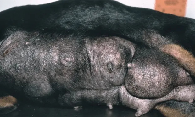
Inflammatory carcinoma, on the other hand, is a diffuse cancer without distinct nodularity, often escapes early diagnosis, and rapidly metastasizes (in days or weeks) to adjacent tissues. Among the divergent types that may be seen are warm, painful diffusely "bruised" mammary glands and fever; lesions that ulcerate to the skin surface and rapidly spread, with involvement along the ventral abdomen and perineum, as in the corgi in Figure 2; dermatologic lesions of the whole ventral abdomen and/or perineum that may be misdiagnosed as contact dermatitis; and diffusely engorged inflamed mammary glands that may be confused with mastitis, as in the Doberman in Figure 1. Although easily diagnosed with a fine-needle aspirate and cytology that shows highly malignant cellular features, the prognosis is grave with minimal or no response to treatment.

