Inappetence & Lethargy in a Cat
Susan Little, DVM, DABVP, Bytown Cat Hospital, Ottawa, Canada
Mischief, a 10-year-old, castrated domestic shorthair cat, was presented for poor appetite and lethargy of 3 days’ duration.
History
Mischief had been a healthy cat with no previous medical problems. He lived indoors and was current on recommended vaccinations. His diet consisted of good-quality dry and canned foods formulated for senior cats.
Physical Examination
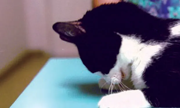
On presentation, Mischief had a BCS of 4/5 and weighed 7.0 kg with no weight loss since previous examination a few months earlier. Generalized weakness and neck ventroflexion were noted (Figure 1). Temperature, pulse, and respirations were normal. Blood pressure was 140/85 mm Hg. All other physical examination findings were normal, including those from neurologic examination.
Figure 1 (above). Generalized muscle weakness and neck ventroflexion are common in cats with profound hypokalemia.
Laboratory Results
CBC and urinalysis results were unremarkable. Blood tests for FeLV antigen and FIV antibody were negative. Serum chemistry panel results are shown (Table).
Ask Yourself
What is the most appropriate initial plan for Mischief?
A. Administer SC fluids, start PO potassium gluconate at 2 mEq q12h, maintain the current diet, reevaluate in 1 week.
B. Administer SC fluids, start PO potassium gluconate at 2 mEq q8h, transition to a renal diet, reevaluate in 1 week.
C. Hospitalize the patient, start IV lactated Ringer’s solution with potassium chloride at 20 mEq/L, monitor serum potassium q12h.
D. Hospitalize the patient, start IV lactated Ringer’s solution with potassium chloride at 40 mEq/L, monitor serum potassium q8h.
Best Answer
D. Hospitalize the patient, start IV lactated Ringer’s solution with potassium chloride at 40 mEq/L, monitor serum potassium q8h.
Profound serum potassium deficits are associated with even lower intracellular potassium concentrations, and aggressive treatment is required. Mischief was started on IV lactated Ringer’s solution at a maintenance fluid rate of 60 mL/kg/24h with the addition of potassium chloride at 40 mEq/L. Serum potassium was monitored q8h. When serum potassium was unchanged after 24 hours, potassium chloride supplementation was increased to 60 mEq/L, and oral potassium gluconate supplementation at 2 mEq q12h was started. Despite these measures, after 3 days of therapy Mischief’s serum potassium remained at less than 3.0 mEq/L.
Further Investigation
The refractory nature of Mischief’s hypokalemia required further investigation to identify its underlying cause. Based on physical examination and initial laboratory data, common causes of hypokalemia were excluded (see Some Common Causes of Hypokalemia).
Some Common Causes of Hypokalemia
Chronic kidney disease
Gastrointestinal disease
Drug-induced (eg, diuretics, insulin)
Postobstructive diuresis after urethral obstruction treatment
Hepatic disease
Endocrinopathies (eg, hyperthyroidism, hyperaldosteronism, hyperadrenocorticism)
Diabetic ketoacidosis
Decreased intake (eg, prolonged anorexia)
Related Article: Feline Hypokalemic Myopathy
Abdominal radiographs were unremarkable. Abdominal ultrasound from a referral hospital revealed a heterogeneous mass in the right adrenal gland measuring approximately 2.2 × 3.4 cm. All other abdominal organs were unremarkable. With the findings of hypokalemia refractory to treatment and a unilateral adrenal mass, a presumptive diagnosis of primary hyperaldosteronism (PHA) was made. While many cases of PHA are associated with both hypertension and hypokalemia, not all patients have both findings, and some patients are presented without clinical signs referable to either hypertension or hypokalemia.1 Mischief fit the typical age for PHA (range, 5–20 years; median age, 13 years).
The adrenal mass was consistent with unilateral adrenal hyperplasia or neoplasia (eg, adenoma, adenocarcinoma, pheochromocytoma). Tests that can support a diagnosis of PHA (when available) include measurement of plasma aldosterone and the plasma aldosterone-to-renin ratio.1,2 Recently, measurement of the urinary aldosterone-to-creatinine ratio after suppression with oral fludrocortisone has been reported as a practical confirmatory test for PHA.3 However, when an adrenal mass is identified (Figure 2), surgical excision and histopathology are necessary for a definitive diagnosis and to plan medical versus surgical treatment.

Ultrasound showing a unilateral adrenal mass in a cat (arrows) with a hypoechoic appearance. Courtesy Dr. Edward Javinsky, Veterinary Medical Consultations
Treatment
Potassium supplementation was continued, and the aldosterone receptor blocker spironolactone (2.5 mg/kg PO q12h) was started. When the serum potassium improved to 3.8 mEq/L, a board-certified surgeon performed a right adrenalectomy. No other abnormalities were found in abdominal organs. Mischief was maintained postoperatively on IV potassium supplementation. Serum potassium concentrations stabilized in the reference range by the third postoperative day when potassium supplementation and spironolactone administration were discontinued.
Related Article: Hyperaldosteronism in Cats
Histologic examination revealed the adrenal mass to be a well-differentiated adenocarcinoma with no evidence of vascular invasion. The mass appeared completely excised.
Follow-up

Mischief at his follow-up examination (3 months postoperatively)
At examination 1 month postoperatively, Mischief had normal serum chemistry panel, CBC, and urinalysis results. Serum potassium was 5.0 mEq/L. Serum chemistry panel was monitored monthly for 3 more months and remained within reference ranges. At last examination, 10 months postoperatively, Mischief was stable with no recurrence of disease.
The Take-Home
Blood pressure should be measured routinely in sick cats.
Hypokalemia that is poorly responsive to treatment should be investigated further.
PHA in cats is likely more common than previously thought and should be included as a diagnostic differential in cats with hypokalemia and/or hypertension after other common causes such as chronic kidney disease are excluded.
The diagnosis of PHA includes imaging the adrenal glands and specific testing such as measurement of plasma aldosterone or the plasma aldosterone-to-renin ratio when available.
Unilateral adrenalectomy is the treatment of choice for confirmed unilateral primary hyperaldosteronism and has high success rates and long-term disease-free times.
1,4
The author thanks the Specialty and Referral Services of Alta Vista Animal Hospital in Ottawa, Ontario, for their key role in Mischief’s diagnosis and treatment.