Human Fetal Ultrasound in the Veterinary Clinic: Is It Safe?
Robert L. Toal, DVM, MS, DACVR, eVet Diagnostics
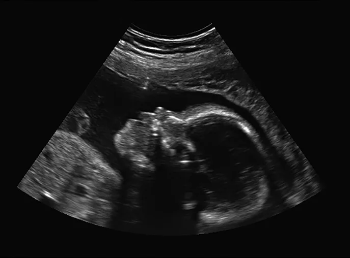
Ultrasound self-examination of the fetus by pregnant veterinary professionals is a growing trend in veterinary medicine. Is this a harmless action, or could there be an unforeseen downside? Let’s take a look at some of the issues.
What is the motivation?
In human medicine, the number of scans performed during pregnancy varies according to physician preference and patient needs. Scans are used to provide information about fetal health, to determine gender, and to obtain printed images of the fetus or fetuses for expectant parents; ultrasound visualization of the pregnancy can also facilitate an emotional connection to the fetus. All of these objectives are important.
Why do some feel the need to do additional scans on their own?
The reasoning includes
Having ready access to the equipment and knowing that the procedure is easy.
Curiosity, especially if expectant parents are excited about identifying gender without having to wait for their next OB/GYN visit.
Worrying about an unexplained change in or lack of fetal movement, which may trigger the need to “take a quick peek” for reassurance that everything is okay.
Enjoying the novelty of seeing the fetus develop over time or the need to have more keepsake images or videos.
So, in summary, convenient access to equipment, curiosity, anxiety, and/or fascination may be key motivators in the decision to self-scan a fetus.
Why do others have no interest in self-scanning?
The reasoning from this group involves
Having trust that their doctor is monitoring fetal progress at appropriate intervals and being comfortable with the level of medical care received.
Being unfamiliar with how to interpret the nuances of self-scanned findings.
Not wishing to introduce unwanted or unfounded concerns.
Satisfaction that the pregnancy is progressing normally.
Interestingly, neither group seems to mention ultrasound safety or potential risks of exposing the fetus to excessive ultrasound imaging.
What are the risks?
For more than 40 years, widespread use of prenatal ultrasound in expectant mothers has not resulted in any recognized adverse fetal effects to date.1
After 1992, improved technology in ultrasound machines demanded more powerful output settings (ie, for better imaging and for Doppler), and the FDA modified output regulations as a result, including allowing a maximum 8 times higher acoustic intensity than that allowed before ultrasound was used for fetal examinations. But, no long-term fetal research studies were conducted during that era.2
Currently, it is recommended that sonographers use the Output Display Standard (ODS)3 as a safety reference guide to limit exposure while trying to obtain the best images during fetal examinations. At times, however, obtaining a better image may create greater risk for the fetus.
Image quality versus fetal risk: How does ODS work?
Ultrasound mechanical wave energy is converted into heat absorbed by the body and the fetus. Higher beam energy and more heat absorption increase the risks for cellular damage.
The ODS for modern ultrasound units displays numbers that are a crude measure of risk due to beam energy effects, including the thermal index (TI) for heat and the mechanical index (MI) for mechanical effects.
These numbers are calculated by the machine and change with
Probe frequency
Mode (B-mode or Doppler)
Harmonic imaging
Width of beam for color flow
Dwell time
Power
All of these parameters affect image quality; adjustments can improve image quality but may increase beam energy and thus fetal risk.
The ODS can include 3 TIs: 1) one for soft tissue (TIS), 2) one for bone at focus (TIB), and 3) one for bone at the surface (TIC), such as the skull.
Bone has a relatively high absorption and heats quickly, while soft tissue heats less efficiently. Thus, fetal trimester and extent of bone development dictate potential heat generation and absorption.2,4
For the MI parameter, several mechanisms of cellular damage attributed to beam energy and mechanical interactions with tissues and/or cells have been documented.2,5
What safety issues and risks are associated with self-scanning?
In general, TI and MI values of <1 are considered safe, although the maximum MI value for the eye remains at 0.23 and many suggest using an MI of <0.4, especially when near gas bodies (eg, lung, bowel) because of potential for cavitation injury (ie, microbubbles rapidly expand and collapse, causing cell damage).<sup2,6,7 sup>
The adjustable power control for most veterinary units is set at 100%, which is the default setting. This means that the beam has maximum acoustic energy all the time. Obstetric examiners often keep the power setting lower to decrease the TI/MI calculations and promote fetal safety.
Duration of the probe use also influences how much acoustic energy is delivered to the fetus. Long scan times or fixed probe positions that linger can deliver more acoustic energy per volume of tissue and therefore are less desirable than well-planned methodical examination by a professional sonographer.2,5 With ultrasound self-examinations, the user may not be familiar with operation protocols (eg, adjusting parameters that would affect TI and MI levels) or may not realize that efficient use of the probe can be key in minimizing fetal exposure.
What is the Principle of Prudent Use?
Although the radiation safety concept of ALARA (As Low As Reasonably Achievable) may be applicable for professional sonographers, in my opinion, the Principle of Prudent Use may be more appropriate here, especially if untrained veterinary staff are performing self-examinations that expose the fetus to potential high-risk energy sources. This principle questions the wisdom of nonmedical scanning, excessive scanning of any patient, scanning a fetus for sales demonstration purposes, and similar generally nonessential uses (eg, keepsake videos).
So, the bottom line is: If you do self-examinations at your veterinary clinic, you may be placing the fetus at risk, especially if you do not know how to properly use the equipment. Instead, the Principle of Prudent Use suggests that you let your OB/GYN group perform fetal ultrasound as directed by your physician.
What do you think?
MI = mechanical index, ODS = output display standard, TI = thermal index