How to Interpret Dental Radiographs
Full-mouth dental radiographs are rapidly becoming standard during dental procedures, as they routinely identify pathology that might not be evident at examination. Dental radiographs can be important for small animal patients that do not consistently demonstrate oral discomfort and pain. This gallery of both normal and abnormal dental radiographs shows various ways in which radiographs can provide insights for diagnosis.
Related Article: Dental Radiography of the Maxillary 4th Premolar, Simplified

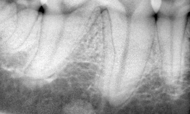
Figure 1. Focal idiopathic Osteosclerosis
These radiographic densities, like this one ventral to the right mandibular first molar (tooth 409), are characterized by bone sclerosis and well-defined borders of varying shape. These lesions are incidental and generally clinically insignificant.
Related Article: Digital Dental Radiology
BRETT BECKMAN, DVM, FAVD, DAVDC & DAAPM, owns and operates a teaching center in Punta Gorda, Florida, dedicated to internationally advancing the educational needs of veterinarians and veterinary technicians in the fields of dentistry and oral surgery. He also accepts dentistry and oral surgery cases in Atlanta, Georgia; Orlando, Florida; and Punta Gorda, Florida. In 2010 Dr. Beckman was acknowledged for his role in dental education with the distinguished Fellow of the Year award from the Academy of Veterinary Dentistry and the Miltex Award for Excellence in Veterinary Dentistry. He earned his degree at Mississippi State University.