Gingival Hyperplasia
Profile
Definition
Gingival hyperplasia is defined as an enlargement of the gingiva that is noninflammatory, produced by factors other than local irritation, and the result of an increased number of cells.1 Because gingival hyperplasia denotes a specific histologic diagnosis, clinicians must rule out other causes of gingival enlargement with biopsies of affected tissues.2 A common example is gingival enlargement caused by local irritation or inflammation.
Signalment
Species. Seen more often in dogs than cats.
Breed predilection. Great Danes, collies, dalmatians, and Doberman pinschers; boxers appear to be overrepresented.3
Related Article: Common Oral Tumors in Dogs and Cats
Causes
Idiopathic
Breed predisposition
Medications
Pathophysiology
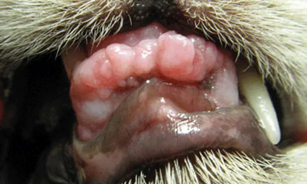
Drug-induced gingival hyperplasia appears to be associated with alteration of calcium influx in gingival tissue. The exact mechanism of action has yet to be determined.4 Calcium antagonism may play a role in aldosterone synthesis by increasing testosterone levels.5 Studies have shown an increase in gingival hyperplasia associated with testosterone injections and a decrease in hyperplasia in castrated dogs receiving oxodipine.6 In patients receiving cyclosporine, transglutaminase levels are decreased in gingival tissue when calcium is not readily available, thereby decreasing apoptosis7 (Figure 1). In feline studies, cells resembling fibroblasts were stimulated to proliferate with the administration of phenytoin; this process resulted in gingival enlargement.8

Gingival hyperplasia in a cat taking cyclosporine for feline lymphocytic-plasmacytic gingivostomatitis.
Signs
Physical examination reveals solitary, multiple, or generalized enlargement of attached gingiva. Inflammation may be secondary to periodontitis but is not responsible for primary enlargement in gingival hyperplasia (see Definition). Bleeding upon probing is a good indicator of periodontal involvement. Tissue is generally firm to the touch. Proliferation may become excessive and gingiva may resemble an oral mass, resulting in mobility upon manipulation.
Diagnosis
Definitive Diagnosis
Definitive diagnosis can be made only by biopsy and histopathologic examination. Gingival hyperplasia is noninflammatory, but periodontal disease can alter this condition clinically and histologically.
Differential Diagnosis
Gingival hyperplasia must be differentiated from other forms of gingival enlargement. Acute and chronic inflammation, generally associated with periodontal disease, can cause generalized or focal gingival enlargement, which may then cause a hyperplastic appearance of the gingiva.


Extensive gingival hyperplasia in a dog (A); the same patient after gingivoplasty was performed with a 15-scalpel blade and a 12-fluted carbide burr (B)
Medications
Medications in the following 3 categories share the mechanism of calcium antagonism, which is thought to play a role in the hyperplastic process.
Immunosuppressants (especially cyclosporine): Have been known to produce gingival hyperplasia.9 This condition has recently been recognized more frequently due to use of cyclosporine therapy for treating atopy in dogs and cats.
Calcium-channel blockers: Shown to produce gingival hyperplasia in dogs treated for cardiac disease.10
Anticonvulsants: Produce gingival hyperplasia in cats and dogs.11,12
Systemic
Although multiple human diseases have been associated with gingival enlargement, systemic associations in dogs and cats have not been adequately documented in the literature.
Neoplasia
Any benign or malignant oral mass is a gingival enlargement that may resemble gingival hyperplasia. Biopsy must be done in these cases. Dental radiography becomes important in differentiating malignant oral masses from hyperplasia. Productive tumors of bone and cysts may cause the appearance of hyperplasia by expanding beneath the attached gingiva. A similar condition is commonly seen in cats with expansion of buccal bone secondary to periodontal disease or root resorption associated with tooth resorptive disease. Gingival hyperplasia should not be confused with proliferative gingival lesions associated with feline lymphocytic plasmacytic gingivostomatitis or tooth resorption.
Treatment
Medical
In 1 study, folate decreased gingival hyperplasia in humans and cats taking the anticonvulsant phenytoin.13 The condition often resolves simply by withdrawing medication, but this solution is often not feasible.14
Surgical
Surgical correction provides definitive pseudopocket removal and reestablishes normal gingival contour (Figure 2). Bulk removal of tissue is accomplished with scalpel excision. Gingivoplasty using a 12-fluted burr provides accurate contour (Figure 3).

12-fluted carbide burr on a high-speed dental handpiece
The pocket depths and landmarks for excision are located with the aid of a periodontal probe and needle to provide bleeding points in the gingiva as a guide to excision.
The periodontal probe is inserted into the pocket to measure depth. It is then removed and placed on the adjacent gingival epithelium.
A needle then marks the pocket depth 2 mm coronal to the actual depth of the pocket.15 This ensures a final sulcus of 2 mm.
A bevel incision is then made to this level at an angle of 45°.
Once gingivoplasty removes the bulk of the hyperplastic tissue, a 12-fluted carbide burr (which I prefer) or a diamond burr is used to provide precise gingivoplasty to approximate the original contour of the gingiva.
Electrosurgery and laser have been used to provide both bulk removal and contouring, but only individuals with extensive experience should consider use of those methods in lieu of the method described.
Client Education
Gingival hyperplasia and gingival overgrowth result in pseudopockets that trap debris and plaque, commonly resulting in periodontal pocketing.
Treatment involves recreating normal anatomy surgically; however, gingival hyperplasia not associated with any specific cause, such as that seen in idiopathic breed-related hyperplasia, will recur in most cases.
The condition usually recurs many months to years later, but clients should be aware that repeated gingivectomy or gingivoplasty may be required.
Medications
Analgesic management is essential when gingivectomy or gingivoplasty is chosen to treat gingival hyperplasia. Premedication with analgesics is commonly used before anesthesia when surgical manipulation of the oral cavity is imminent. Regional nerve blocks are needed to ensure immediate postoperative comfort and, most important, optimal anesthetic safety. Postoperative analgesics are indicated for 4 to 6 days.
Follow-Up
Patient Monitoring
Although postoperative complications are not likely when scalpel gingivectomy and burr gingivoplasty are used, 7-day rechecks are advised. If the operator chooses to use the more invasive techniques, follow-up should be at 7-day intervals for 3 to 4 weeks to ensure that there are no signs of thermal damage.
Complications
Complications generally arise only when laser surgery and electrosurgery are used for gingivectomy or gingivoplasty. Proper settings, operator experience, and precise technique are paramount if these methods are chosen. Thermal necrosis of gingival tissue, bone, and teeth can result when these methods are used improperly (Figure 4).

Thermal necrosis in a patient treated for gingival hyperplasia using radiosurgery. Generalized bone, soft tissue, and pulp necrosis resulted in multiple extractions.
Future Follow-Up
Annual or biannual examinations can be used to monitor for recurrence of hyperplastic tissue. Need for repeated surgery should be determined according to the extent of the changes.
In General
Prognosis
Prognosis for a reasonable interval of clinical resolution is excellent. Many cases do recur, however, and surgical intervention may be required periodically to control the condition.
Cost Considerations
General anesthesia and analgesia are required for surgical management. Severity of the condition varies from focal, isolated involvement to generalized hyperplasia. Therefore, the cost of therapy ranges from $$$ to $$$$$.
Treatment at a Glance
Medical
If possible, discontinue calcium-channel blockers, anticonvulsants, or immunosuppressants.
Consider using folate supplementation, which has been shown to decrease drug-induced gingival hyperplasia in humans.
Surgical
Apply regional nerve blocks.
Measure pocket depths.
Create bleeding pockets with a needle.
Remove bulk gingiva with a scalpel.
Contour remaining gingiva with a 12-fluted carbide burr or diamond burr.
Postoperative
Provide analgesic management.
Recheck in 7 days.
Use annual or biannual examinations to monitor for recurrence.