Feline Tooth Resorption—Extraction of Retained Premolar Root
R. Michael Peak, DVM, DAVDC, Tamp Bay Veterinary Specialists, Largo, Florida
The incidence of feline tooth resorption (formerly termed resorptive lesions) has been estimated at 30% to 70%.1 Because some experts believe that there are 2 different and distinct patterns of root resorption, each treated differently, dental radiographs are a crucial part of diagnosis.2 The importance of full-dentition radiography has also been reported.3
Types of Resorption & Treatment
As suggested by DuPont and DeBowes, 2 feline tooth roots with resorption can be classified as having type 1 or type 2 patterns of resorption.
Roots with type 1 resorption are identified radiographically as having a focal area of radiolucency in the crown or root, coupled with evidence of obvious root structure, with normal radiopacity and a well-defined periodontal ligament space.
Roots with type 2 resorption have indistinct delineations between alveolar bone and root structure, with generalized loss of radiopacity and an indistinct periodontal ligament space.
Roots showing type 1 resorption should be extracted, but some debate exists about proper treatment for type 2. One approach for the latter is to visually examine the alveolar crestal bone and remove any clinically evident roots. If none are evident or extraction is not possible, this approach calls for intentionally retaining the resorbing segment and suturing the gingiva over it. This approach should not be used if there is any evidence of endodontic disease, advanced periodontitis, or feline stomatitis.
Step-by-Step: Extraction of Retained Premolar Root
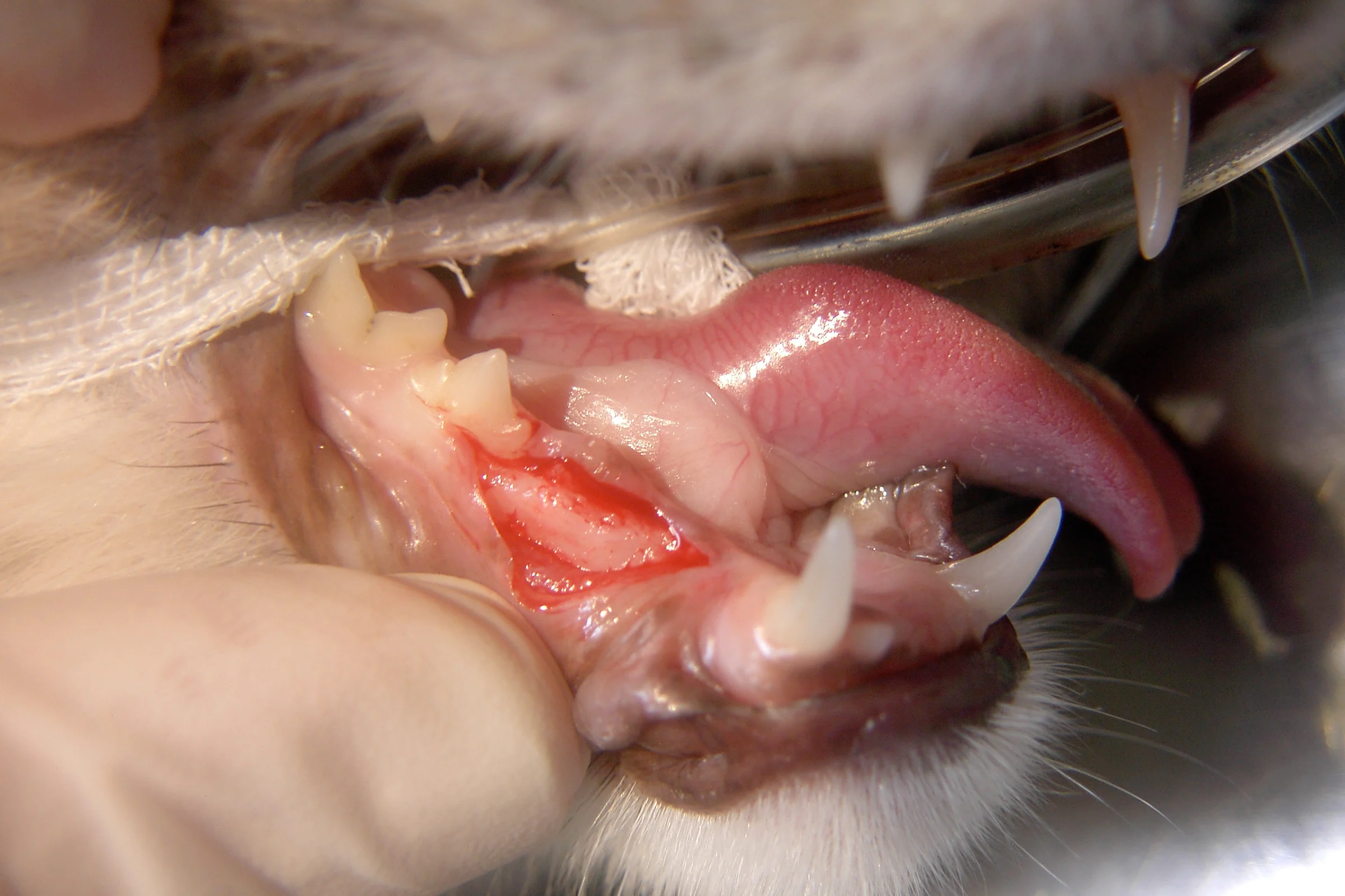
The patient in the following figures was presented for possible tooth resorption. During anesthetized oral examination, the crown of the right mandibular third premolar (#407) was noted to be missing; however, there was inflammation over the mesial aspect of the area where this tooth would have been. An intraoral radiograph revealed an obvious, clearly defined mesial root and an indistinct remnant of the distal root, which was undergoing type 2 root resorption.
What You Will Need
Molt #2 periosteal elevator
Taper diamond burr
Small (#1/4 round) burr
Winged elevator
Extraction forceps or thumb forceps
Absorbable suture
Step 1
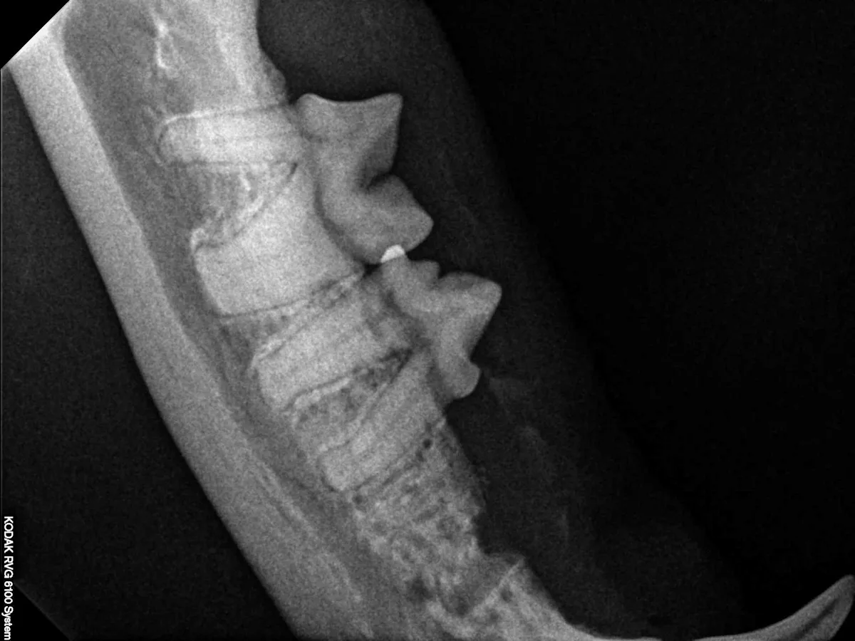
Intraoral radiograph of #407 shows a clearly defined intact mesial root (arrow) and ill-defined distal root with type 2 resorption. The clinical lesion of gingivitis is noted over the retained mesial root.
Step 2
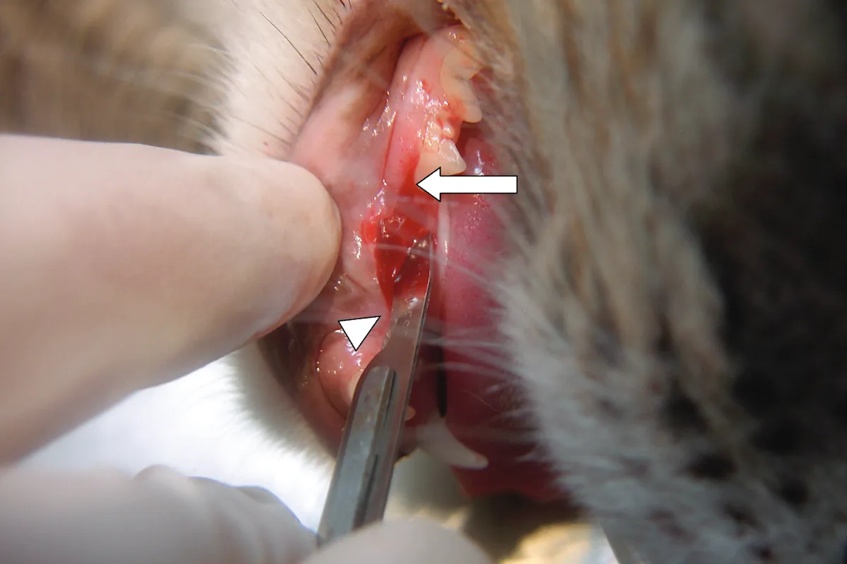
After a right mandibular nerve block, an incision is made from the mesiobuccal line angle (arrow) of the mandibular right fourth premolar (#408) to the attachment of the right mandibular frenulum (arrowhead). This location allows full examination of both the distal and mesial roots of #407. As an alternative, and if needed, a vertical releasing incision can be made mesial to #408 or along the mesiobuccal line angle of #408.
Author Insight
An incision from the mesiobuccal line of the mandibular 4th premolar to the attachment of the mandibular frenulum allows full examination of both the distal and mesial roots.
Step 3
The gingiva is elevated on the vestibular as well as the lingual aspect by using a Molt #2 periosteal elevator to expose the crestal alveolar bone.
Author Insight
The Molt #2 periosteal elevator is used to expose the crestal alveolar bone. A taper diamond burr is used to smooth the crestal bone and reevaluate for evidence of root structure.

Step 4
The crestal alveolar bone is inspected for clinical evidence of root structure. The mesial root can be seen. The distal root is not evident.

Step 5
A taper diamond burr is used to smooth the crestal bone and reevaluate for any clinical evidence of root structure. Retraction of the lip or gingival margins may be necessary while performing this osteoplasty.

Step 6
A small (#1/4 round) burr is used to “outline” the periodontal ligament space of the mesial root, working circumferentially around the root structure 2 to 3 mm apically.4
Author Insight
Excavation around the retained mesial root allows for easy insertion of a dental elevator.

Step 7
Excavation has been completed around the retained mesial root. This allows easy insertion of a dental elevator alongside it, with minimal apical pressure applied to the root.

Step 8
A winged elevator is used to gently place slow, steady rotational pressure on all sides of the retained root segment. Gentle pressure is held for 10 to 20 seconds, and the elevator position is then moved. Pressure continues until the root is sufficiently displaced for its removal with an extraction forceps or thumb forceps.

Step 9
The retained root segment has now been elevated and is loose enough for removal.
Author Insight
Gentle pressure is held for 10 to 20 seconds on all sides of the retained root segment until the root is sufficiently displaced for removal.

Step 10
The mesial root after extraction: the area of the distal root is again examined for evidence of clinical roots. If necessary, the alveolar bone is smoothed with the diamond burr. Before closure, the surgical site is irrigated and examined for debris.

Step 11
An postoperative intraoral dental radiograph shows that the mesial root has been removed and the alveolar crestal bone over the distal root has been smoothed.
Step 12
Absorbable suture is used to close the gingiva.

Outcome
This patient was discharged and prescribed a 5-day course of oral analgesics. The owner was instructed to feed a softened diet for 7 to 10 days. No complications were noted when the site was reevaluated for healing 14 days later.
Advantages & Complications
The advantage of this technique is that it allows good visualization of the retained root segment while creating an area for dental elevator placement without the need for excessive apical pressure on the retained root segment. Excessive pressure could displace the root segment into the mandibular canal.
Potential complications include root fracture (if that occurs, the procedure can be applied further apically along the root), hemorrhage from disruption of the mandibular artery (if entered), and iatrogenic mandibular fracture. Although no published studies report on the incidence of such complications, the rate should be low when the above extraction techniques are applied.