Abdominal Distension & Third Eyelid Protrusion in a Young Cat
Katherine Clarke, BVSc, MRCVS, BSAVA, PGCertSAM, DECVIM‑CA, Davies Veterinary Specialists, Hertfordshire, United Kingdom
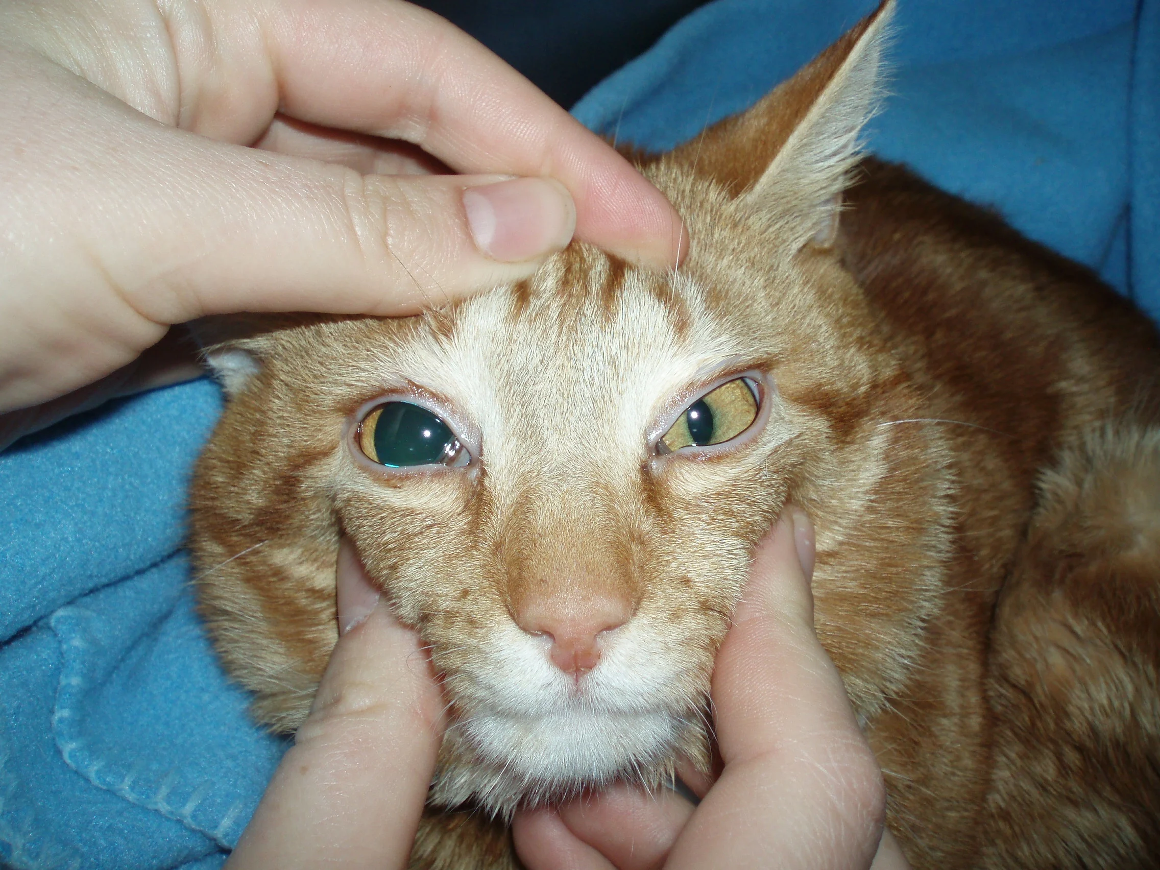
Marmalade, an 18-month-old, neutered male, indoor/outdoor domestic shorthair cat, was presented with a short but progressive history of vomiting, anorexia, third eyelid protrusion, abdominal distension, and depression. Because Marmalade urinated and defecated outside, the owner was unable to provide information about fecal consistency or urination habits. There was no previous medical history; parasite preventives and vaccinations were current.
Physical Examination
On physical examination, Marmalade was quiet but responsive. Bilateral third eyelid protrusion and bilateral mydriasis were present. Mucous membranes were dry/tacky with bilateral crusting around the external nares (ie, xeromycteria). Cardiac auscultation identified relative bradycardia (heart rate, 115 bpm) with a sinus rhythm, no audible heart murmur, and synchronous pulses. Abdominal palpation identified caudal organomegaly suggestive of bladder enlargement, resulting in abdominal distension. He was normothermic; BCS was normal.
Differential Diagnoses
Dysautonomia was considered based on clinical signs (ie, vomiting, anorexia, bilateral nictitans protrusion and mydriasis, tacky mucous membranes, nasal crusting, bradycardia, large urinary bladder) and apparent multisystem dysfunction; however, dysautonomia is largely a diagnosis of exclusion. Methodical investigations are therefore needed to exclude other differential diagnoses (eg, primary GI disturbance causing anorexia, dehydration, presumptive vagally mediated bradycardia due to abdominal pathology, vomiting-induced rhinitis, mydriasis secondary to situational fear of the clinic).
Diagnostics
IV fluid therapy was started immediately to correct any dehydration.
CBC and serum chemistry profile results were within reference intervals, including normal potassium, absence of azotemia, and normal protein values. Blood pressure measurement, ECG, and echocardiogram were performed to better understand the bradycardia; results indicated a structurally normal heart, sinus bradycardia, and normotension. An atropine response test (0.04 mg/kg SC) was performed, but there was no response (ECG closely monitored for 60 minutes with no change in rate or rhythm observed), indicating a lack of appropriate sympathetic input to the heart and supporting the diagnosis of dysautonomia.
Direct and consensual pupillary light responses were bilaterally negative; a Schirmer tear test (STT) was 0 mm/min bilaterally. A pilocarpine response test (0.05%) resulted in rapid miosis of the pupil, which is consistent with dysautonomia (Figure 1). Absence of tear production can explain the nasal crusting.
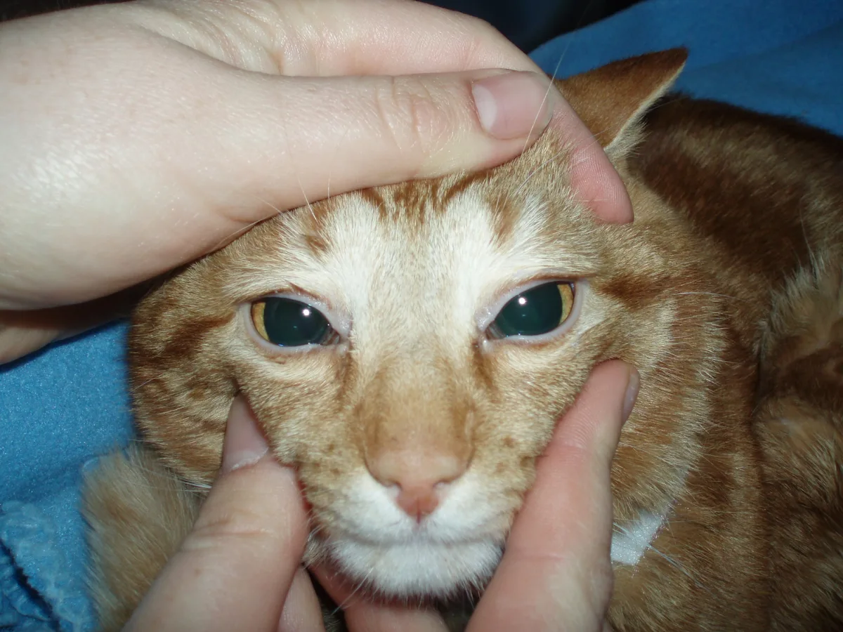
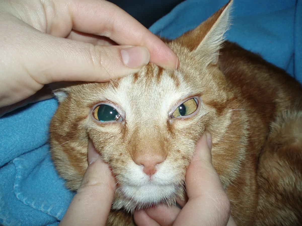
FIGURE 1 Marmalade before (A) and after (B) application of 0.05% pilocarpine to the patient's left eye
Thoracic and abdominal radiography were performed with the patient under sedation with butorphanol (0.25 mg/kg), medetomidine (5 micrograms/kg), and ketamine (2 mg/kg) IV, as well as flow-by oxygen provided via mask, to assess for further evidence of dysautonomia (eg, urine retention, GI ileus, megaesophagus) and secondary associated complications (eg, aspiration pneumonia) and exclude other differentials for vomiting. No evidence of megaesophagus was found, and a very large urinary bladder was confirmed (Figure 2). Abdominal ultrasonography was also performed; generalized intestinal ileus and a very large urinary bladder were identified without evidence of cystitis (normal bladder wall, no evidence of neoplasia or urolithiasis). A urinary catheter was easily passed into the bladder, thus excluding urethral obstruction as a cause for the enlarged bladder. Urinalysis revealed well-concentrated urine (urine specific gravity, 1.055) and mild, microscopic hematuria (20 RBCs/high-power field); results were otherwise unremarkable.
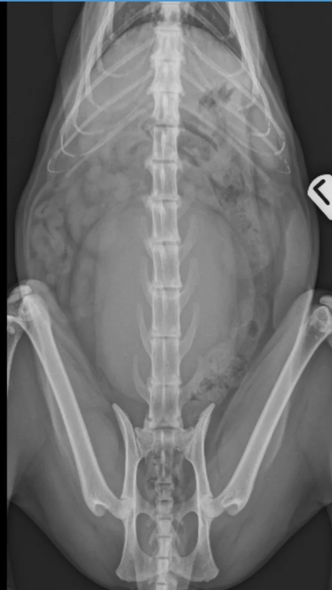
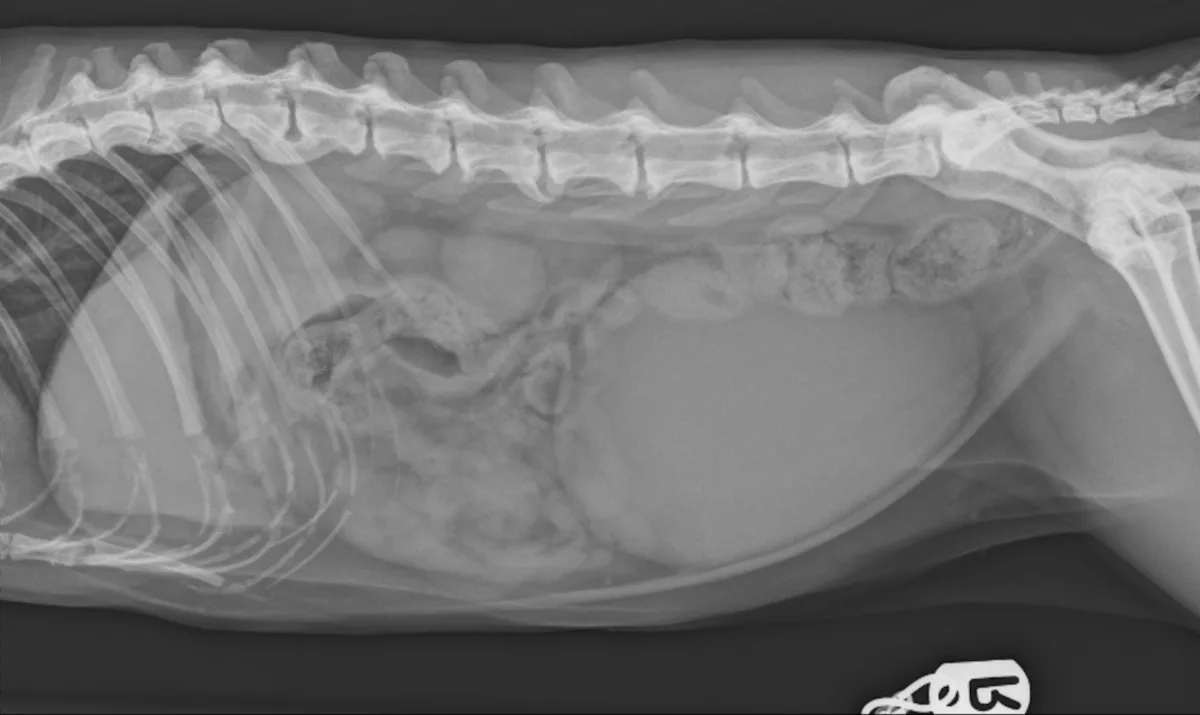
FIGURE 2 Dorsoventral (left) and right lateral (right) abdominal radiographs demonstrating marked urinary bladder distension and moderate accumulation of feces in the descending colon
Diagnosis: Presumptive Dysautonomia
Treatment
Medical management consisted of maropitant (1 mg/kg IV every 24 hours) for nausea, ongoing IV fluid therapy, and suturing the urinary catheter in place to manage urinary retention and allow the detrusor muscle to recover from overdistension (see Tables 1 and 2). Ocular lubrication was regularly applied because of the absence of tear production. A low dose of buprenorphine (0.01 mg/kg IV every 6-8 hours) was administered while the urinary catheter was in place to ensure patient comfort.
The patient remained anorexic, and an esophageal feeding tube was placed. Feeding was initiated via a liquid recovery diet at one-third of the resting energy requirement. Feeding initially went well, but the patient started to regurgitate. A conscious, focal ultrasound identified moderate to severe gastric distension consistent with gastric ileus. Metoclopramide (2 mg/kg/day IV CRI) was started; cisapride (2.5 mg PO/cat every 8 hours) was added because metoclopramide alone failed to encourage gastric emptying. Gastric distension improved, and tube feeding was tolerated. The volume of food was gradually increased to the full resting energy requirement.
After 3 days, the urinary catheter was removed, and urination behaviors and bladder size were closely monitored. Dysuria (ie, straining to urinate without passing urine) was observed on multiple occasions. The urinary bladder became distended, resulting in mild overflow urinary incontinence. A urinary catheter was thus placed with the patient under sedation, and the bladder was fully emptied. Prazosin (1 mg/cat PO every 8 hours) was started to help the urethra relax; bethanechol (2.5 mg/cat PO every 8 hours) was administered to help the detrusor muscle contract during urine voiding. The urinary catheter was removed after 24 hours, and urination behaviors improved.
Outcome
Marmalade was hospitalized for 10 days. He had begun eating small amounts of food, and urination behaviors were near normal. Mild constipation that developed during the hospital stay was resolved with lactulose (2 mL/kg every 12 hours). Prazosin (1 mg/cat PO every 8 hours), bethanechol (2.5 mg/cat PO every 8 hours), maropitant (2 mg/kg PO every 24 hours), lactulose (2 mL/kg PO every 12 hours), ocular lubrication, and cisapride (2.5 mg/cat PO every 8 hours) were prescribed, and an esophageal feeding tube was in place on discharge.
How to Avoid Overwhelming Pet Owners With Information
Owners bringing in pets with wide-ranging signs, as seen with dysautonomia, may already feel overwhelmed. Avoid inundating owners with information and help them retain key concepts with these tips.
Prepare the owner by letting them know you are about to share a lot of information.
Split information into chunks, and be strategic about what is shared when.
At the first appointment, only share information that is critical. Save noncritical information for future conversations.
Use handouts, and empathize with the owner about the amount of information to be shared.
Reading the handout to the owner and highlighting important information can be helpful.
Provide links to helpful videos.
For more on helping education stick, check out this article on Avoiding Overwhelming Clients With Information
After 6 weeks, bethanechol and prazosin were tapered down over 2 weeks and stopped without a change in urination behaviors. By this stage, the patient did not require maropitant to maintain his appetite, and fecal consistency had declined, thus lactulose had stopped resulting in normal fecal consistency and volume. After 2 months of consistent eating, the esophageal feeding tube was removed. An attempt was made to stop cisapride, but hyporexia and reflux behaviors resulted. Pupil size returned to normal; however, alacrima and bradycardia persisted. Cisapride was continued for 12 months before successful tapering and discontinuation.
Two years after the initial diagnosis, the patient made a full recovery except for continued ocular lubricants.
Discussion
Dysautonomia describes the clinical manifestation of autonomic nervous system (ANS) dysfunction. The ANS regulates involuntary physiologic processes (eg, heart rate, GI motility, salivation, pupillary responses, vascular tone).1 Pathologically, dysautonomia is characterized primarily by degeneration of autonomic neurons.1 Abnormalities associated with dysautonomia include depression, regurgitation, constipation, anorexia or hyporexia, dysphagia, dysuria, vomiting, dilated and unresponsive pupils, third eyelid protrusion, dry nares and mucosa, reduced lacrimation, orthostatic hypotension, and bradycardia.1 A selection (rarely all) of these signs are typically present.
The etiology of dysautonomia in veterinary species remains uncertain, although a possible association with Clostridium botulinum neurotoxin has been described in horses and cats.2-4 Diagnosis typically relies on a combination of clinical signs, exclusion of other causes, and confirmatory tests (eg, pharmacologic pupillary response).
Pilocarpine is a direct-acting parasympathomimetic drug that activates cholinergic receptors within the iris sphincter muscle. When applied topically to the eye, pilocarpine induces miosis by stimulating muscarinic receptors. In patients with dysautonomia, degeneration of the postganglionic neurons results in enhanced sensitivity of the denervated muscle to cholinergic drugs.5 This hypersensitivity enables a response to a diluted solution of pilocarpine.
Reduced or absent tear production is a recognized feature of dysautonomia in cats and dogs6; however, low STT values have been reported in clinically normal cats.1 What constitutes a low reading can be contentious, with some publications stating STT of ≈12 mm (±5) over 60 seconds is normal,7 while others state readings <9 mm over 60 seconds should be classified as abnormal.8 Thus, STT is not always a helpful tool for diagnosis of dysautonomia in cats.
Treatment focuses on clinical signs, is often prolonged, and can be challenging. Management of regurgitation, consequential recurrent aspiration pneumonia, and urinary retention that requires repeated catheterization or prolonged urinary catheter management can be especially difficult. Aspiration pneumonia should be confirmed prior to instituting antibiotic therapy; pneumonitis alone does not require antibiotics.
Veterinary literature reports a poor to grave prognosis for affected patients,1,6,9 with euthanasia often elected due to poor treatment response. Recovery is possible in some cases but often takes several months and requires significant commitment from the owner.
Take Home Messages
Dysautonomia should be considered early in patients with evidence of ANS dysfunction.
If bradycardia is present, an atropine response test can be useful. Atropine, a direct-acting parasympatholytic agent, blocks the action of acetylcholine at the muscarinic receptors in the parasympathetic nervous system. Under normal circumstances, IV or IM atropine causes an increase in heart rate as the sympathetic nervous system becomes dominant. Dysautonomia can cause a loss of sympathetic nervous system innervation to the heart; consequently, no change in heart rate is seen in response to atropine administration.
If inappropriate mydriasis is present, a pilocarpine response test can help with diagnosis. Pilocarpine is a direct-acting parasympathomimetic drug that activates cholinergic receptors within the iris sphincter muscle and induces miosis by stimulating muscarinic receptors when applied topically to the eye.
Loss of tear production is common; STT should thus be performed in all suspected cases.
Etiology of dysautonomia is unknown. Treatment is supportive and focused on clinical signs.
Recovery is possible in some cases, but the recovery period is protracted.