Source
Heinze CS, Miles JE, Gawor J, Kortegaard HE. A cross-sectional radiographic study on the prevalence and distribution of dentigerous cysts in unerupted teeth in adult dogs. J Small Anim Pract. 2026;67(5):422-431. doi:10.1111/jsap.70067
Research Note
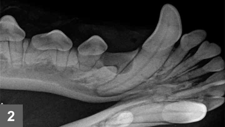
Dentigerous cysts (ie, odontogenic cysts that form around the crown of uninterrupted or partially erupted teeth) result from abnormalities in tooth development, grow slowly, and can affect adjacent cortical bone but are rarely associated with pain or clinical signs other than the absence of ≥1 tooth.
This study investigated the prevalence and distribution of dentigerous cysts in adult dogs with unerupted teeth. Dental radiographs from ≈13,000 records identified 206 (1.58%) dogs with 1 or more unerupted teeth (n = 285). Of these teeth, 95 (33.3%) were associated with dentigerous cysts. The most frequently unerupted tooth was the mandibular first premolar, followed by the mandibular third molar and the central mandibular incisor. With the exception of the mandibular second incisor, the prevalence of cysts per site across all tooth types was <50%. Brachycephalic dogs were more likely to experience cyst occurrence than nonbrachycephalic or crossbreed dogs, and neutered males were more likely to experience cyst occurrence than intact females.
The authors concluded that the percentage (33.3%) of unerupted teeth with dentigerous cysts suggests that monitoring unerupted teeth for radiographic evidence of cyst development may be an acceptable minimally invasive management alternative to prophylactic extraction of unerupted teeth.
You are reading Research Notes, a research summary resource presented by Clinician's Brief. Clinician's Brief does not conduct primary research.