To Cut or Not To Cut: Lameness in a Labradoodle
Case: 14-month-old castrated Labradoodle
History: 4 weeks’ duration of left thoracic limb lameness; lameness worsened with exercise but improved mildly after administration of carprofen at 2mg/kg Q12h.
Related Article: Osteochondritis Dissecans of the Shoulder
Physical Examination: The patient demonstrated grade 1/4 lameness in the left thoracic limb, exhibiting mild pain on left shoulder extension. No pain was detected in the right thoracic limb or neck. Primary differentials for shoulder lameness in a young dog include panosteitis, osteochondrosis, biceps/supraspinatus/infraspinatus tendinopathy, and collateral ligament desmitis.
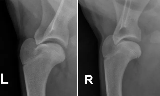
Shoulder Radiographs: Lateral views of both the left and right (Figures above) shoulders were performed. Craniocaudal views were also obtained (not shown).
Related Article: Anthroscopy in Small Animals
Figures above. Lateral views of the left and right shoulders.

Radiographic Findings: Subtle subchondral lucencies were identified along the caudal margin of both humeral heads, supporting osteochondrosis (OCD). No panosteitis or other abnormalities are detected.
_
Figures 2A and 2B. Same images as Figures 1A and 1B, but with arrows pointing to the subchondral defects._
Should this dog go to surgery?
Arthroscopy of both shoulders was recommended.

Comments: CT was performed to confirm the lesions. Figures 3A and 3B are images reformatted into the sagittal plane, showing the distinctly abnormal ossification of the subchondral bone in both humeral heads with an osteochondral fragment (Figure 3A, arrows) adjacent to the left humeral head (osteochondritis dissecans).
Conservative therapy (eg, rest and NSAIDs) may provide short-term relief, but long-term resolution of the lameness requires arthroscopy or arthrotomy with removal of any cartilage fragments and curettage of the abnormal bone/cartilage. Prognosis for normal limb function with shoulder OCD is good with surgical intervention; after surgery, most dogs become sound within 2 months. OCD has a hereditary component, so owners should be advised not to breed affected dogs.
Figure 3A. The reformatted sagittal CT image of the left shoulder; the arrows are pointing to the osteochondral fragment.
Figure 3B. The reformatted sagittal CT image of the right shoulder; the arrowhead is pointing to the subchondral defect.
Outcome: The patient underwent bilateral shoulder arthroscopy to debride the subchondral lesions and to remove the fragment in the left shoulder. His lameness resolved by the 2-week postoperative checkup.
Suggested Reading
1. Computed tomographic diagnosis of non-gastrointestinal foreign bodies in dogs. Jones JC, Ober CP. JAAHA 43:99–111, 2007.2. Radiographic, computed tomographic, and ultrasonographic findings with migrating intrathoracic grass awns in dogs and cats. Schultz RM, Zwingenberger A. Vet Radiol Ultrasound 49:249–255, 2008.3. Comparison of ultrasound, computed tomography, and magnetic resonance imaging in detection of acute wooden foreign bodies in the canine manus. Ober CP, Jones JC, Larson MM, et al. Vet Radiol Ultrasound 49:411–418, 2008.