Complications of Severe Tick Infestation
History
Bear, an 8-year-old neutered male cocker spaniel, and Yoto, a 4-year-old intact male Pyrenees cross, were presented with weakness and anorexia. Bear was presented in mid-July with severe weakness. Bear lives outdoors and was current with his vaccines. According to his owners, he was given a monthly internal parasite-control product (specific product not identified) but did not receive routine flea and tick control.
Yoto also lives outdoors. He was presented in late September for lethargy of 1 week’s duration with several instances of vomiting. His vaccinations were reported as “current” by his owner, but records were not available. Yoto was not on a monthly heartworm and intestinal parasite control product or external parasiticide.
Related Article: Tick-Borne Infectious Disease in U.S. Dogs
Examination

Both Bear and Yoto were nonambulatory at presentation with evidence of hypovolemic shock, including tachycardia (heart rate 140 and 150 beats per minute, respectively) and tachypnea (30 and 36 breaths per minute, respectively), poor peripheral pulses, and pale pink mucus membranes. No icterus was noted in either dog.
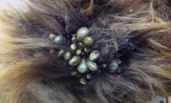
Figure 1. Ticks attached to Bear at presentation. Note cluster of engorged, feeding female ticks surrounded by smaller feeding male ticks and immature ticks.

No free fluid was found in the thorax or abdomen with targeted ultrasound of both patients. It was difficult to find areas of skin that were not occupied by ticks on both dogs (Figures 1 and 2). All ticks were initially identified by the attending clinician as nymphal and adult Rhipicephalus sanguineus, the brown dog tick. This identification was confirmed by a boarded parasitologist by microscopic examination and comparison to standard keys.
Related Article: Practical Guide to Tick-Borne Disease
Figure 2. Feeding adult and immature ticks clustered on the muzzle of Yoto. The skin and surrounding fur are darkened by tick frass and serous exudate from associated tissue damage.
Whole blood was collected for complete blood count. Pertinent laboratory values obtained from each dog are provided in Table.
Ask Yourself
1. What are the differentials for this anemia?2. Do these patients require blood transfusions?3. Could tick infestation be a cause of the anemia, and how should severe tick infestations be treated?4. What is the life cycle of Rhipicephalus sanguineus?
Diagnosis
Tick exsanguination
Treatment
Major cross-matching was performed, and both patients were administered whole blood transfusions to replace blood volume and red blood cells. Yoto’s shock was deemed severe enough, based on degree of anemia, tachycardia, and profound weakness, that he was also administered boluses of synthetic colloid and crystalloid fluids to help stabilize his circulatory volume while whole blood was being prepared from resident, prescreened donors and administered. Packed cell volume (PCV) in both cases initially improved after the first transfusion but began to decline quickly, and both dogs required second transfusions within 24 hours.
Several days of bathing and gentle manipulation were required to remove the bulk of the ticks. The immediate area around the kennels required continuous monitoring to remove and kill ticks as they detached from the patients.
Ticks were initially killed using a topical pyrethrin spray (Vet-Kem Ovitrol Plus, VPL.com) and the dogs were bathed in warm water with a pyrethrin shampoo (Mycodex, VPL.com) to attempt to manually remove as many ticks as could be gently dislodged, taking care to avoid hypothermia. Once patients were dry, fipronil spray (Frontline, merial.com) was applied to as many of the affected areas as possible. Several days of bathing and gentle manipulation were required to remove the bulk of the ticks.
The immediate area around the kennels required continuous monitoring to remove and kill ticks as they detached from the patients. Both dogs were treated with cefpodoxime (10mg/kg q24h for 7 days) for concurrent dermatitis at tick attachment sites and with doxycycline (5 mg/kg q12h for 21 days) as a prophylactic treatment for potential tick-associated rickettsial diseases transmitted by R sanguineus, including Ehrlichia canis, Anaplasma platys, and Rickettsia rickettsii.
Both patients were in a debilitated state and were simultaneously affected by severe anemia, tissue hypoxia, hypoproteinemia, and severe dermatitis.
Outcome
Both dogs survived and were returned home within 5 days after each received 2 whole blood transfusions. A single antiinflammatory dose of an intermediate-acting corticosteroid (dexamethasone, 0.1 mg/kg IV) was used to help control inflammation and the resulting continued loss of protein associated with serous exudate at the tick bites. Because anemia was the result of blood loss, single doses of iron (15 mg/kg IM iron dextran) and cyanocobalamin (25 µg/kg SC) were administered. The patients were discharged on a high-quality, high-energy diet with 2 weeks of oral iron supplementation (75 mg q24) per dog.
Related Article: Diffuse Lower Motor Neuron Dysfunction in Dogs
PCV = packed cell volume, WBC = white blood cell
AARON HERNDON, DVM, DACVIM (SAIM), is a lecturer in small-animal medicine at Oklahoma State University Center for Veterinary Health Sciences. A graduate of Texas A&M University, Dr. Herndon is completing the requirements to earn a doctorate of philosophy in veterinary biomedical sciences.
SUSAN LITTLE, DVM, PhD, DACVM (Parasitology), is a regents professor and Krull-Ewing endowed chair in veterinary parasitology at Oklahoma State University, where she oversees a research program focused on ticks and tick-borne diseases. A past-president of the American Association of Veterinary Parasitologists, Dr. Little is co-director of the National Center for Veterinary Parasitology and the president of the Companion Animal Parasite Council. She frequently presents at the annual North American Veterinary Community Conference.