Cloacal Prolapse in a Bearded Dragon
Spike, a 3-year-old Australian bearded dragon of undetermined sex, was presented for “something coming out of his hind end.”
History. The prolapse had been present for 2 days. The owner tried soaking the tissue in a sugar solution, which had been recommended on an Internet forum, but no improvement was noted. Spike is housed with one other bearded dragon of similar size. Proper heating, lighting, and diet were provided.
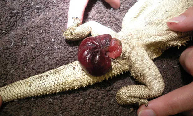
Physical Examination. Spike was bright, alert, and responsive. Body condition was adequate. Bones were firm, and no abdominal masses were palpated. Protruding from the vent was 3 cm of edematous pink tissue, traumatized but viable.
Imaging. The whole-body radiograph was unremarkable.

Figure 1: Traumatized edematous tissue protruding from the vent of a bearded dragon

Figure 2: The radiograph of the bearded dragon did not help determine the cause of the cloacal prolapse
Ask Yourself...
Which of the following is the best initial assessment of this case?A. Prolapse of the gastrointestinal tractB. Prolapse of the bladderC. Prolapse of the oviductD. Prolapse of the hemipenes
Correct Answer:A. Prolapse of the gastrointestinal tract
One of the most important parts of treating prolapses in reptiles is identifying the tissue or organ that has prolapsed. Reptiles have a single cloacal opening called the vent. The intestines, urinary bladder (in species where it is present), and reproductive system all exit from this opening. Any one of these organs can prolapse.

Diagnosis. Close examination may help evaluate which tissue has prolapsed because each will have a unique appearance. Unfortunately, edema and trauma may make identification difficult.
Intestinal or colon prolapses are usually identified by the smooth tubular appearance of the tissue. Sometimes feces may be observed inside the prolapsed tissue.
Oviductal tissue appears similar to intestine, but it has longitudinal striations and there are no feces inside.-Bladder tissue is thin walled and often fluid filled.-Hemipenes are solid tissue and originate from the lateral caudal portion of the vent.
Determining the sex will also help rule out 1 reproductive organ over another. In this case, the prominent femoral pores along the thighs helped determine that Spike was a male.
Causes. Causes of cloacal prolapse include intestinal parasites, bacterial infections, enteritis, constipation or fecal impaction, dystocia, cystic calculi, neoplasia, or trauma. Intestinal stasis secondary to hypocalcemia has also been implicated. Fecal examination for parasites, blood analysis, and abdominal radiography may be required to determine the cause.
Treatment. Clean the exposed tissue with saline or diluted chlorhexidine solution, removing any superficial crust that may have formed. Edema can be reduced by hypertonic saline, sugar solutions or glycerin, and gentle pressure. Once the swelling is reduced, use a lubricated cotton-tipped swab or blunt probe to return the tissue to its normal orientation. In some cases, sedation or general anesthesia may be helpful. Devitalized tissue may require resection.
After the prolapse is reduced, place a purse-string suture around the cloaca or place transverse cloacal sutures. (Use a local anesthetic if general anesthesia was not used for reduction of the prolapse.) This will decrease the size of the opening to prevent repeat tissue prolapse, but will leave just enough space for urine to pass.
Antibiotics and antiinflammatories are warranted. Keep the pet well hydrated; however, do not feed in order to prevent production of more feces and defecation through the sutured vent. Remove the sutures in 3 to 5 days and resume feeding.
Prognosis. If the cause of the prolapse is not identified and resolved, there is a good chance that the prolapse will happen again. Prognosis depends on the underlying cause, condition ofthe patient, and viability and duration of the prolapsed tissue.
Take-Home Message
-Cloacal prolapses can include the gastrointestinal tract, urinary bladder, or reproductive organs.
-Cloacal prolapses usually respond well to reduction of the tissue and limiting the size of the cloacal opening for 3 to 5 days.
-Without determining and treating the original cause of the cloacal prolapse, the prolapse will most likely return.