Guide to Cheyletiellosis
Darren Berger, DVM, DACVD, Iowa State University
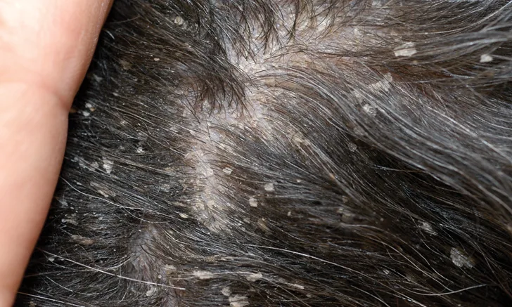
A 10-year-old spayed Scottish terrier with cheyletiellosis demonstrating excessive, large scale formation. Photo courtesy of J.O. Noxon, DVM, DACVIM, Iowa State University
Cheyletiellosis, also known as walking dandruff, is an uncommon, contagious dermatosis caused by an infestation of the surface-dwelling Cheyletiella spp mite.
Cheyletiellosis may occur in dogs (caused by C yasguri), cats (caused by C blakei), or rabbits (caused by C parasitivorax) and can also cause a transient infestation in humans that come in contact with pets carrying the mites. An increased incidence of mites may be observed in immunocompromised patients, in geographic regions where routine flea prevention is not practiced, or following exposure to high-volume housing situations (eg, catteries, breeding facilities).
Cheyletiella spp mites have a standard life cycle of egg, larva, nymph, and adult that can be completed in roughly 21 days. Mite life stages can be identified via direct examination of collected debris using a powerful magnifying lens or via microscopic examination of superficial skin scrapings, acetate tape impressions, and fecal flotation specimens. Cheyletiella spp mites are obligate parasites, as larvae, nymph, and adult male mites die soon after leaving the host; however, adult female mites are more robust and may survive up to 10 days off the host.1
Clinical Signs
Clinical signs are highly variable, and subclinical carriers may be encountered. The most common clinical findings include mild-to-intense pruritus, excessive scaling (particularly over the dorsum), and erythema (Figures 1 and 2). In addition, cats may be presented with barbering alopecia or miliary dermatitis.1

Figure 2
The dorsum of a 3-year-old neutered male cocker spaniel with mild erythema of the skin and lightly adherent scale formation secondary to Cheyletiella spp infestation
Diagnosis
Diagnosis is confirmed via visualization of the mite or ova, which may be difficult to recover from some patients with low-grade infestations. Adult mites can be easily identified by the presence of prominent hooks on their accessory mouthparts (Figures 3 and 4). Cheyletiella spp ova appear similar to louse eggs but are nonoperculated, smaller, and loosely attached to hairs (Figures 5 and 6).
Cheyletiellosis should be considered a differential diagnosis in patients with pruritus and excessive scaling; other ectoparasites, poor nutrition, intestinal parasitism, and primary seborrhea would also be considered differential diagnoses. In addition, Cheyletiella spp infestation should be eliminated as a potential cause in any patient presented for evaluation of a suspected allergic hypersensitivity (eg, atopy, food allergy).
Treatment
No licensed products are indicated specifically for the treatment of cheyletiellosis. Therapeutic protocol and medication selection primarily depend on the species of the animal affected and clinician preference. Most acaricidal flea preventive products and lime sulfur are effective, provided all in-contact animals are treated, the patient is treated for 6 weeks to disrupt the parasite’s life cycle, and conventional environmental treatment—similar to what is recommended for flea infestation—is performed to prevent reinfestation.