Chest Tube Placement
Lisa L. Powell, DVM, DACVECC, BluePearl Veterinary Partners, Eden Prairie, Minnesota
Chest tube placement (ie, tube thoracostomy) plays an important role in the management of severe pleural space disease. Chest tubes are indicated for managing unresolving pneumothorax, pyothorax, persistent pleural space disease requiring multiple thoracenteses, and postoperative thoracotomies.
Chest tube placement can often be performed under controlled conditions, with the patient sedated and intubated to allow regulation of breathing. Sometimes, however, a chest tube needs to be placed emergently or in a nonintubated patient and requires firm knowledge of the procedure.
In a noncrisis, adequate general anesthesia should be induced with placement of an endotracheal tube, allowing the patient to breathe on its own. If oxygenation is inadequate because of severe pleural space disease, gentle positive-pressure ventilation can be applied. In addition, an incision can be made into the chest cavity to relieve tension pneumothorax, with care to support ventilation, as the normal negative pressure within the chest is no longer present.
Related Article: Detection & Treatment of Pneumothorax
Related Article: How to Place a Chest Tube
Related Article: Thoracocentesis
Step-by-Step: Chest Tube Placement
What You Will Need
Thoracostomy tube (comparable in size to the diameter of the patient’s mainstem bronchus as determined by lateral radiography)
Sterile surgical pack (ie, blade, blade handle, needle drivers, suture scissors, drapes, straight Kelly hemostats)
Suture material for skin closure and to secure tube (3-0 nylon for the skin,
polydioxanone for tube)
C-clamp for tube occlusion
Christmas tree adapter, zip-tie, and 3-way stopcock for the end of the chest tube

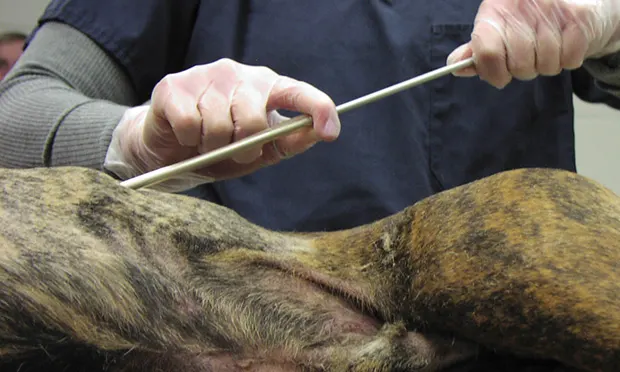
STEP 1.
Once the patient is intubated and stable under anesthesia with adequate oxygen saturation readings, position the patient in lateral recumbency (either side) and widely clip the lateral thorax (from caudal to the thoracic limb to the caudal aspect of the last rib). Use an appropriately sized, sterile chest tube to measure the distance in centimeters from the anticipated skin incision (tenth intercostal space) to the ventral region just caudal to the thoracic limb. Place the chest tube on a sterile field, and prepare the thorax aseptically.
Author Insight
Maintain sterility while measuring the tube (ie, do not touch the thorax with the tube).
Tube Thoracostomy: International Insight
The “pound method” of placing a thoracostomy tube is no longer recommended, as underlying tissue damage and tube misplacement is more likely to occur.
The patient should be anesthetized and intubated, if possible, before placing the chest tube for complete control of respirations when the chest is excised.
Carmalt hemostats should not be used to clamp off the thoracostomy tube once it is placed in the chest cavity. Traumatic damage to the tube may occur, causing air leakage into the chest cavity after placement; an atraumatic C-clamp should be used instead.
Once the chest tube is removed, the surgical site should be left to heal via second intention, and not sutured closed, as air leakage may occur under the skin for a period of time after the chest tube has been removed.
This article is part of the WSAVA Global Edition of Clinician's Brief.