Canine Facial Dermatoses Challenge
Skin conditions involving the face of dogs can look similar but have very different causes. Use the pictures in addition to clues from the signalment to match each picture with the definitive diagnosis. In a clinical setting, you would have the benefit of many additional pieces of information, such as physical examination, history, and diagnostic tests-this exercise does not suggest that diagnosis should ever be made on appearance alone, and it is unlikely that the reader will make the correct diagnosis in all cases.
Empirical therapy based on a tentative diagnosis should not be attempted in many of these diseases. Immunosuppressive therapy is strongly contraindicated when infectious disease has not been ruled out.

1. 4-month-old chow chow: Demodicosis
This puppy has generalized demodicosis with secondary pyoderma. Clinically, this might be difficult to differentiate from juvenile cellulitis (puppy strangles). However, juvenile cellulitis on the face initially manifests as acute swelling of the eyelids, lips, and muzzle, which later progresses to papules and pustules involving the lips, muzzle, chin, bridge of the nose, and periocular area. This puppy has confluent involvement of the haired region of the face; thus, deep skin scrapings should be part of the diagnostic plan.

2. 4-year-old Great Dane: Scabies
This adult dog has extensive self-trauma to its head and face. This degree of self-trauma first suggests Sarcoptes scabiei, and treatment for scabies should be done even if skin scrapings are negative.
Related Article: Sarcoptic Mange

3. 2-year-old rottweiler: Vitiligo
Depigmentation without inflammation on the muzzle, lips, and nose pad in this breed are highly suggestive of vitiligo. The disorder is most often seen in rottweilers, Belgian tervurens, and Doberman pinschers. Biopsy would confirm. Leukotrichia or poliosis sometimes accompanies this condition.

4. 3-month-old golden retriever: Juvenile cellulitis
Scrapings should be done for Demodex. The mucocutaneous location of the lesions, the swelling and cracking of affected areas, and the age of the pup suggest this condition. One would expect physical findings to include enlarged peripheral lymph nodes. These patients can be depressed and febrile. Retrievers and dachshunds seem to be overrepresented.

5. 8-year-old Labrador mix: Pemphigus foliaceus
This dog has a symmetrical crusting/scaling disorder that involves the bridge of the nose, the periocular region, and perhaps the pinnae. Depigmentation of the nose has occurred. Crusting and scaling were probably preceded by some type of primary eruption, which could be dermatophytosis; however, the nose pad involvement, facial symmetry, and age of the dog more likely suggest pemphigus, neoplasia, or superficial necrolytic dermatitis. Diagnosis would involve cytologic evaluation, biopsy, and a thorough medical examination of the dog’s metabolic status.
Related Article: Canine Pemphigus Foliaceus

6. 2-year-old smooth-coated collie: Dermatomyositis
The lesions are focal areas of hyperpigmentation, alopecia, and atrophy. Muscle atrophy over the bridge of the nose is apparent. Collies are one of the breeds predisposed to this disorder, and the appearance of the lesions is highly suggestive. Diagnosis would be confirmed through skin biopsies and EMG or muscle biopsy.

7. 5-year-old akita: Uveodermatologic syndrome (Vogt-Koyanagi-Harada)
The breed, the location of the lesions, and the obvious uveitis give the diagnosis away!

8. 1-year-old German shepherd: Insect hypersensitivity
The lesions are present only on the dorsum of the nose in the lightly haired areas and appear to be somewhat nodular. They could be neoplastic (although the age of the patient makes that diagnosis unlikely) or inflammatory. Inflammatory nodules, which appear on the nose rather suddenly, usually result from insect bites, although they may result from fungal or bacterial infection.

9. 3-year-old bloodhound: Atopic dermatitis
This dog has evidence of chronic self-trauma and erythema; he is also licking his feet and scratching at the trunk. This case of atopic dermatitis is complicated by secondary Malassezia infection.

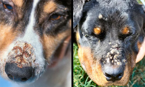
10. 1-year-old rottweiler: Dermatophytosis
This adult dog has focal crusts, small nodules, and accompanying moderate alopecia. Focal alopecia without nodules is found on the ears and the forehead. This inflammation is associated with a Microsporum gypseum infection from the soil and resembles insect bites or other infections, including nasal pyoderma.

11. 10-year-old Welsh corgi: Superficial necrolytic dermatitis
Other names for superficial necrolytic dermatitis are necrolytic migratory erythema, metabolic epidermal necrosis, and hepatocutaneous syndrome. This geriatric dog has crusting and cracking on the nose and around the eyes. The disorder could be pemphigus foliaceus or epitheliotropic lymphoma (Mycosis fungoides ), but the patient has larger crusts than usually occur after the pustules of pemphigus foliaceus. Biopsies and a thorough medical workup would help with diagnosis. This dog has underlying liver disease. Pancreatic neoplasia, especially glucagonoma, has been associated with this condition.
12. 6-year-old spaniel mix: Discoid lupus erythematosus (cutaneous lupus)

The condition is confined to the nose pad and is characterized by inflammation, a slate-gray depigmentation, and loss of cobblestone appearance. The loss of cobblestone appearance can occur with other facial conditions, such as neoplasia or other autoimmune disorders.