Anesthetic Protocols for Brachycephalic Dogs
Tasha McNerney, CVT, CVPP, VTS (Anesthesia & Analgesia) , Veterinary Anesthesia Nerds Glenside, Pennsylvania
Brachycephalic dogs are becoming more popular as pets,1 which means veterinary nurses are more likely to be asked to anesthetize these dogs in practice. Brachycephalic dogs have a relatively broad, short skull, usually with the breadth at least 80% of the length.2 They often have anatomic abnormalities (eg, stenotic nares, elongated soft palate, hypoplastic trachea, laryngeal collapse, everted laryngeal saccules), known as brachycephalic syndrome, which can cause upper airway obstruction and mandate the use of special protocols when administering anesthesia. (See Common Abnormalities.)
Anesthetic Premedication & Oxygenation
Sedation
Sedation is an important component of anesthetic premedication. α-2 agonists (eg, dexmedetomidine), phenothiazines (eg, acepromazine), or benzodiazepines (eg, diazepam, midazolam) are often administered. Excitable or nervous brachycephalic patients may be prescribed higher doses of premedication sedation agents; however, deep sedation can cause excessive relaxation of upper airway muscles and worsen airway obstruction.3 Lower doses should be administered in these patients unless they are aggressive or dangerous.
Analgesia
Always provide analgesia for surgical procedures. Opioids, which are used most frequently for perioperative analgesia, are not contraindicated in brachycephalic patients, despite the potential for respiratory depression, depending on the dose.4 Opioids commonly used for perioperative analgesia include morphine, hydromorphone, oxymorphone, fentanyl, buprenorphine, and butorphanol. Unlike phenothiazines and benzodiazepines, α-2 agonists such as dexmedetomidine also provide analgesia. When combined with other medications in the anesthetic premedication protocol, dexmedetomidine may provide sufficient analgesia and muscle relaxation for minor surgical procedures.5
Consideration of Anticholinergic Agents
Anticholinergic agents, which block muscarinic receptors, are typically included in anesthetic premedication protocols to minimize parasympathetic effects of anesthesia such as bradycardia, bronchoconstriction, and excessive saliva formation.6 Brachycephalic breeds often have higher vagal tone than other breeds and can become bradycardic,4 so anticholinergic agents such as glycopyrrolate may be administered to elevate heart rate. Routine use is not recommended, but anti-cholinergics can be given to decrease secretions and reduce the likelihood of aspiration pneumonia on a case-by-case basis. They are contraindicated in patients with certain cardiac diseases (eg, mitral insufficiency) in which an elevated heart rate is harmful and should be used with caution in patients with myocardial oxygen balance issues.7
Preoxygenation
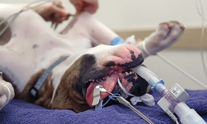
Preoxygenate brachycephalic patients for 10 to 15 minutes following administration of anesthetic premedication agents. This can increase fraction of inspired oxygen (FiO2), which may improve partial pressure of oxygen (PaO2).4 Administer induction agents intravenously rather than by facemask, which is not recommended for brachycephalic patients because it can increase stress. The edge of the facemask can also damage the patient’s cornea, and use of rapidly metabolized induction agents is preferred in these patients.8 Intubation will require a small-diameter endotracheal tube and use of a laryngoscope because excessive tissue in the pharynx may reduce the visibility of the laryngeal opening.
Anesthesia Maintenance
Maintain anesthesia with an inhalant such as isoflurane or sevoflurane in 100% oxygen. Sevoflurane is less soluble than isoflurane and allows patients to recover more quickly,9 an important consideration for brachycephalic patients. During the intraoperative period, use of a multiparameter monitor can provide constant information about the patient’s status, including electrical activity of the heart, peripheral oxygen saturation (SpO2), end tidal CO2 (ETCO2), temperature, and blood pressure. ETCO2 readings indicate the patient’s ability to ventilate by measuring CO2 in exhaled respiratory gases and are closely correlated with arterial carbon dioxide (PaCO2) under normal conditions. Normal canine and feline ETCO2 is 35-45 mm Hg.10
Recovery
The recovery period is as important as the anesthetic period and requires equally vigilant patient monitoring. Recovery should be as smooth and stress-free as possible for all patients but is especially important in brachycephalic breeds because of their respiratory compromise. As for all patients, the veterinarian will prescribe appropriate postoperative analgesic agents based on the level of pain anticipated from the surgery. Note that acepromazine has no analgesic properties. Brachycephalic patients can sometimes desaturate when in recovery, so a portable pulse oximeter should be used to monitor hemoglobin saturation.
Place postoperative brachycephalic patients in sternal recumbency with the head slightly elevated. Avoid overly aggressive initial stimulation, which may trigger movement and/or swallowing followed by a relapse into unconsciousness when the stimulation is removed. Make sure additional induction agents and endotracheal tubes are available if an airway obstruction occurs and reintubation is needed. Recovering brachycephalic patients in an oxygen chamber is advisable; however, if an oxygen chamber is unavailable at the practice, nasal oxygen via a red rubber catheter can be an alternative.11 A nasopharyngeal tube can be placed and connected directly to an oxygen source to allow delivery of oxygen to the oral cavity during recovery.11
Brachycephalic Breeds12,13
Brachycephalic breeds need extra care because they are unable to cool themselves sufficiently, as they have difficulty breathing, especially in hot conditions or during excessive exercise. The soft tissue in the palate obscures the trachea and prevents air from flowing over the tongue, which is how most dogs cool themselves. Brachycephalic breeds include Boston terrier, boxer, bull mastiff, Brussels griffon, Dogue de Bordeaux, English bulldog, French bulldog, Lhasa apso, Pekingese, pug, and shih tzu.
Common Abnormalities
The most common abnormalities observed in brachycephalic breeds are:
Stenotic nares: A condition in which the nostrils are too narrow and sometimes collapse inward during inhalation, making it difficult for the patient to breathe through the nose
Hypoplastic trachea: A condition in which the diameter of the trachea (ie, windpipe) is smaller than normal
Elongated soft palate: A condition in which the soft palate is too long and its tip protrudes into the airway, interfering with inspiration of air into the lungs
Long-Term Hospitalization
The ventilation status of brachycephalic patients requiring long-term hospitalization should be assessed frequently. Use portable monitors to measure SpO2 and arterial blood gas samples when available, and keep oxygen readily available. Maintain body temperature as close to normal range as possible.
If brachycephalic patients show signs of stress during hospitalization, the veterinarian and veterinary nurse should work together to determine if the patient is painful or anxious. For pain, appropriate analgesics should be administered. To manage anxiety, administer medications such as trazodone or a microdose infusion of dexmedetomidine.
Conclusion
Brachycephalic dogs have anatomic abnormalities that require careful airway and respiration monitoring during and after procedures requiring anesthesia. Use of appropriate anesthetic premedication helps ensure smooth induction, and vigilant monitoring during all stages of anesthesia and recovery reduces potential complications, making working with these patients less challenging and more rewarding.
This article originally appeared in the March 2017 issue of Veterinary Team Brief.