Allergic Dermatitis in a Puppy: More Than Meets the Ear

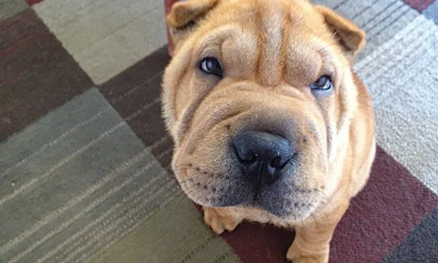
Beignet, a 3-month-old, 10-kg, castrated Chinese shar-pei, presented for a 1-month history of scratching his ears, neck, and chest along with excessive licking of his forelimbs.
History
On a scale of 0 to 10, with 0 representing minimal pruritus and 10 representing severe, constant pruritic behaviors, the client reported a score of 7.
The patient’s diet consisted of a commercial chicken and rice–based dry food with occasional table scraps. He received monthly heartworm prevention (ivermectin 0.006 mg/kg PO) but not flea control. Vaccines were up-to-date. Beignet was primarily an indoor dog with outdoor access to groomed, grassy areas. The other nonrelated pet in the home, a 10-year-old spayed shar-pei, was not pruritic.
Related Article: Identifying Causes of Otitis Externa
Physical Examination
The patient was bright, alert, and responsive. His heart rate, temperature, and respiratory rate were within normal limits. Cardiac and thoracic auscultation and abdominal palpation were unremarkable.
Figure 1. On presentation, stenosis of the horizontal canal with purulent exudate was noted during otoscopic examination in a 3-month-old shar-pei.

Cutaneous examination revealed diffuse alopecia, erythema, hyperpigmentation, papules, crusts, and mild lichenification of the ventral neck and chest. The distal forelimbs were diffusely erythematous with mild alopecia.
The ear canals were palpably firm and pliable, and pain was elicited during external manipulation. The pinnal–pedal reflex was negative. Otoscopic examination revealed bilateral moderate erythema and severe stenosis of the horizontal ear canals with purulent exudate (Figure 1).
Diagnostics
Deep skin scrapings of the ventral neck and chest were negative for Demodex spp. Superficial skin scrapings of the neck and chest were negative for Sarcoptes scabiei. Acetate tape cytologic samples of the ventral neck and chest revealed 10 to 20 rods and 5 to 10 Malassezia spp per high power field (hpf).
Cytologic samples from beneath crusts on the ventral neck and chest showed neutrophils and 3 to 10 intracellular and extracellular cocci/hpf. Cytology of the right ear was positive for 20 to 50 rods/hpf and 10 to 20 cocci/hpf. Cytology of the left ear was positive for 10 to 20 cocci/hpf. Mite preparation of ear exudate was negative.
Diagnosis
Cutaneous adverse food reaction
Preliminary Diagnosis
A preliminary diagnosis of allergic dermatitis and allergic otitis externa was made based on the lack of overt causes of otitis (eg, ticks, mites, tumor) as well as the findings of superficial staphylococcal folliculitis, Malassezia spp dermatitis and bacterial overgrowth of the neck, and bacterial and yeast otitis externa with stenosis.
Treatment
The following treatment plan was recommended for Beignet’s bacterial and yeast dermatitis:
Cephalexin 25 mg/kg PO q12h until recheck
Miconazole (2%)–chlorhexidine gluconate (2%) shampoo twice weekly
Ketoconazole (1%)–chlorhexidine gluconate (2%)–acetic acid (2%) wipe applied to the neck and chest once daily
The following treatment plan was recommended for otitis externa with stenosis:
Prednisolone 1 mg/kg PO q24h until recheck. The client was informed that prednisolone might result in temporary shrinking of the muzzle and wrinkles, because of reduced hyaluronic acid production. The characteristic skin wrinkles and muzzle appearance of the shar-pei breed are secondary to cutaneous mucinosis.
Enrofloxacin (0.5% w/v)–silver sulfadiazine otic (1.0% w/v) emulsion 0.3 mL applied q12h for 1 week, then q48h until recheck.
Fluocinolone acetonide (0.01%)–dimethyl sulfoxide (DMSO; 60%) otic solution: 5 drops q48h.
A restrictive diet trial was instituted with a prescription dry venison and potato diet, approved for growing puppies. The canned formula was provided for hiding pills, and treats were limited to cooked potato.
Related Article: Diagnosis & Management of Otitis
A monthly topical spot-on containing moxidectin (2.5%) and imidacloprid (10%) was recommended for heartworm control to replace the flavored chewable. Flavored medications and supplements should be replaced with non-oral or non-flavored formulations during an elimination diet trial because many products include animal- and plant-derived proteins that may be allergenic.
Figure 2. At the first recheck (2 weeks after initiation of treatment), the dog’s ear was erythematous with mild hyperplasia and ceruminous exudates. The breed-related narrowness of the ear canal in addition to discomfort precluded a complete view of the tympanic membrane without anesthesia.

A recheck was recommended in 10 to 14 days.
Outcome
Recheck 1
Beignet’s pruritus score was 2/10. The owners reported mild polyuria/polydipsia and polyphagia. Crusting and papular dermatitis were nearly resolved. The ventral neck, chest, and distal forelimbs were mildly alopecic and erythematous. The muzzle was approximately 30% reduced in size.
The ears were normal on palpation. Stenosis of the horizontal canal was mild for the breed with mild hyperplasia. Yellow-brown ceruminous exudate and medication were present, preventing complete visualization of the tympanic membranes (Figure 2).
Repeat cytology of the ears revealed occasional rods and cocci. The restrictive diet trial was continued, and cephalexin was continued for 1 additional week. Prednisolone at 0.25 mg/kg PO q48h was continued for 2 additional weeks.
The shampoo and wipes were maintained twice weekly. Flushing the ear canal with a Tris-EDTA product twice weekly was prescribed to remove exudate and enhance the antibacterial effect prior to application of the enrofloxacin–silver sulfadiazine emulsion. Fluocinolone–DMSO otic solution was also continued twice weekly with instructions to discontinue 1 week prior to the next recheck, which was recommended in 4 weeks.
Recheck 2 & Beyond
Figure 3. Eight weeks after initial presentation, the otitis externa had resolved and the tympanic membrane could be visualized.

At the second recheck, 8 weeks after initial presentation, Beignet’s pruritus score was 0. Mild hypotrichosis was present on the neck. The ears were nonpainful and pliable on palpation. The ear canals demonstrated scant erythema. Tympanic membranes were visualized and normal (Figure 3).
Repeat cytology of the ears revealed no etiologic agents.
A tentative diagnosis of cutaneous adverse food reaction was made. However, the seasonality of the pruritus was unknown given the young age of the patient. Therefore, the possibility remained that maintenance of a clinically normal ear upon discontinuation of antiinflammatory therapy may have been the result of a season change in an atopic patient rather than improvement because of the elimination diet.
Re-challenge of the diet elimination trial with the patient’s original diet after week 8 was performed to confirm cutaneous adverse food reaction. Pruritus and otitis returned several days after feeding of the previous diet. The strict diet was resumed and pruritus once again resolved within 2 weeks. No further treatment was required.
hpf = high power field
JENNIFER SCHISSLER PENDERGRAFT, DVM, MS, DACVD, is assistant professor at Colorado State University’s James L. Voss Veterinary Teaching Hospital in Fort Collins, Colorado. Her special interests include cutaneous infectious disease, antimicrobial resistance, infection control practices, clinical immunology, and otology. After completing a 1-year rotating internship in small animal medicine and surgery at Wheat Ridge Animal Hospital in Wheat Ridge, Colorado, she completed a combined master’s degree and dermatology residency at The Ohio State University.