Top 5 Ways to Differentiate Primary Cardiac From Primary Pulmonary Disease in Emergencies
Elke Rudloff, DVM, DACVECC, Lakeshore Veterinary Specialists, Glendale, Wisconsin
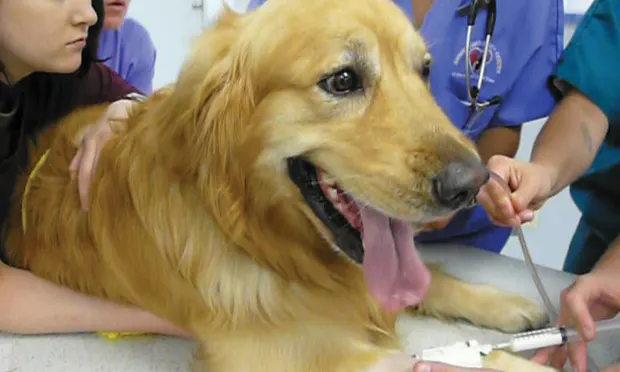
Differentiating primary cardiac from primary pulmonary disease in small animals with respiratory distress can be daunting. To hurriedly force a patient starving for air into a position to take diagnostic radiographs is a misguided and life-threatening maneuver; instead, immediate therapeutic intervention and a thoughtful diagnostic approach are more effective. The initial approach in treating severe respiratory signs is to provide oxygen supplementation and sedation (eg, butorphanol, 0.4 mg/kg IV or IM; midazolam, 0.2 mg/kg IV or IM) to reduce anxiety associated with dyspnea. IV catheter placement, tracheal intubation, assisted ventilation with 100% oxygen, and suctioning of the airway may be necessary before diagnostic evaluation (even in a patient with congestive heart failure [CHF]).
Related Article: Respiratory Distress: Diagnosis & Treatment at a Glance
1. Examination
Perfusion parameters can be altered with both primary cardiac and pulmonary diseases. Mucous membrane color can range from pink to pale pink to cyanotic, regardless of the degree of distress. In most instances, patients with primary cardiac disease have normal to subnormal body temperature, whereas patients with pulmonary disease have normal to increased body temperature.
A key to localizing the origin of a respiratory problem is to identify the breathing pattern (Table 1). When auscultating the thorax, it is best to listen to all lung fields before focusing on heart sounds. In this order, louder heart sounds do not desensitize the examiner from hearing finer changes associated with lung disease. Generalized reduced lung sounds may suggest pleural space occupation with fluid or air. Focal reduction in lung sounds may identify a lobe that is not aerating (eg, torsion). Moist crackles can develop with pulmonary edema from cardiogenic and noncardiogenic causes, as well as with interstitial inflammation. If the crackles are focal, lobar pulmonary disease (eg, aspiration pneumonia, contusion, neoplasia) is more likely. If diffuse moist lung sounds are accompanied by a heart murmur and/or a cardiac dysrhythmia, then pulmonary edema secondary to CHF should be strongly considered. Heart murmurs in cats can be very focal or intermittent, and patience is required when auscultating the heart. Jugular venous distension may also support a primary cardiac problem.
Table 1: Breathing Patterns & Region Affected
*Synchronous breathing describes the chest and abdomen moving together during inhalation and exhalation; asynchronous breathing describes the chest and abdomen moving opposite from each other during inhalation and exhalation.
2. Patient history
There is an increased suspicion for primary cardiac disease if there is historical evidence of heart disease, a known heart murmur, exercise intolerance, and/or coughing without activity. A cough may also be supportive of lower airway disease (eg, tracheobronchial inflammation) as well as heartworm disease. Recent history of anesthesia, vomiting, or regurgitation may prompt a directed examination for aspiration pneumonia. History of trauma or exposure to an anticoagulant drug or toxin may initiate investigation for intrathoracic hemorrhage. Anticoagulant intoxication can cause a variety of cardiopulmonary problems, including pulmonary interstitial hemorrhage, hemothorax, mediastinal hemorrhage, and pericardial effusion—all of which can manifest with respiratory signs. Evidence of rib fractures or other external injuries may accompany pulmonary contusion or hemothorax resulting from trauma. A history of a cough in the dog and cat may prompt evaluation for lower airway disease. A recent prolonged seizure event, brain injury, systemic inflammatory response syndrome, or choking or electrocution may hasten the development of noncardiogenic pulmonary edema.
Related Article: Acute Respiratory Distress: The Blue Patient
3. Ultrasonography
In the acute care setting, thoracic-focused assessment sonography for trauma (TFAST) can be used during immediate evaluation of the patient with respiratory distress—with minimal handling or positioning—to identify the presence of pneumothorax, pleural effusion, pericardial effusion, atrial enlargement, a hypodynamic heart, and diffuse or infiltrative pulmonary changes (Table 2). Although it requires experience and training, ultrasonographic evaluation of the thorax can be completed rapidly and with the same, or increased, degree of sensitivity and specificity as thoracic radiography when identifying pleural space disease and cardiac dysfunction.1,2 A comprehensive echocardiogram is necessary in all cases of suspected cardiac disease to confirm or rule out the presence and type of underlying myocardial disease and guide definitive treatment.
Table 2: Thoracic Ultrasound Changes Associated with Primary Cardiac & Pulmonary Disease Causing Respiratory Distress
4. Thoracic radiography
A number of changes can be identified on thoracic radiographs to help distinguish primary cardiac from primary pulmonary disease; however, radiography should not supercede immediate intervention and stabilization. Information from a comprehensive physical examination and history should be sufficient to initiate reasonable treatment for immediate stabilization before seeking thoracic radiographs. Any pleural space air or fluid should be evacuated before obtaining thoracic radiographs; this not only stabilizes the patient for radiographic positioning but also permits adequate insufflation of the lungs and improves diagnostic quality of lung evaluation. If the patient is intubated and receiving assisted ventilation with oxygen support, then radiographs should be taken during the administration of a gentle inspiratory breath not exceeding 20 cm H2O.
Primary cardiac disease that leads to CHF-related pulmonary edema commonly causes diffuse (perihilar in the dog) or variable (in the cat) interstitial and alveolar patterns. The vertebral heart score (VHS) is an objective method for evaluating the size of the cardiac silhouette (see Steps to Measure VHS). Certain cutoff values are used to determine whether acute respiratory distress is primarily pulmonary or cardiac in origin and to guide initial therapy for stabilization and planning for definitive cardiac or respiratory workup. A VHS <11.4 in the dog can help rule out mitral valve disease–related CHF as a cause for respiratory signs.3 Although an increased VHS can be associated with primary cardiac disease, it is not a specific characteristic in small-breed dogs because of their thoracic conformation.3 In the cat, a left lateral VHS ≤7.9 can reasonably rule out the presence of heart disease.4 Additional radiographic changes associated with primary cardiac disease include left atrial enlargement and pulmonary venous distension (Figures 1 and 2). Pleural effusion can be associated with CHF and primary pulmonary disease. A miliary pattern and/or solitary soft tissue densities in the lungs are indicative of primary pulmonary disease (Figure 3). Hyperinflation of the lung and distinct peribronchial markings are supportive of lower airway disease, although peribronchial markings can be found in the cat with CHF.

Figure 1.
Right lateral thoracic radiograph of a dog that was presented with labored breathing and a moist cough. The patient had a history of a heart murmur, and the breathing pattern was synchronous with increased effort on inhalation. Thoracic auscultation revealed diffuse, soft moist crackles and a 4/6 left systolic heart murmur over the mitral region. Butorphanol and furosemide were administered and the patient was allowed to rest with supplemental oxygen before radiography. Radiographs showed a VHS of 12.5, significant left atrial enlargement (white arrows), and pulmonary venous distension (black arrows) consistent with left-sided heart failure. Mitral valve insufficiency caused by valvular endocardiosis was confirmed with echocardiography.
5. Laboratory testing
A plasma N-terminal pro-B-type natiuretic peptide concentration (NT-proBNP) >1400 pmol/L in the dog and >270 pmol/L in the cat with respiratory difficulty, regardless of the presence of a heart murmur, is more likely to be found with primary cardiac problems than with primary respiratory problems.5 A blood sample should not be collected until the patient can tolerate restraint. Ultimately, this screening measure takes time to obtain results and does not replace the need for echocardiography to confirm primary cardiac disease.
Closing thoughts
Mixed cardiac and pulmonary disease can exist in small animals with respiratory distress. For example, a dog may be presented with a history of mitral valve insufficiency but have clinical and radiographic evidence of aspiration pneumonia following a dental prophylaxis; or, a cat may present with a heart murmur and evidence of lower airway disease based on breathing pattern and thoracic radiographs (Figure 4; see image gallery above).
If a primary cardiac problem cannot be ruled out based on history and examination, or there are conflicting signs as to whether respiratory distress is primarily cardiac or pulmonary in origin, a dose of a diuretic (eg, furosemide at 2 mg/kg IV or IM) can be given pending diagnostic imaging. There should be little harm in administering a single dose in a patient that is not significantly dehydrated, and it could be of benefit if there is CHF.
Steps to Measure VHS

Measure the long axis (LA) of the heart from top of the left atrium to the tip of the apex of the ventricle (blue arrow).
Measure the short axis (SA) of the heart at the widest part of the left and right heart chambers at the level of the coronary groove (red arrow).
Transpose the LA and SA dimensions onto the vertebral column and record the corresponding number of vertebrae (V), starting at the cranial edge of T4 moving caudally.
Take the sum of the SA and LA vertebral measurements to find the VHS.5. In this patient, the VHS = 9.5.
CHF = congestive heart failure