Vestibular Disease: 3 Case Studies
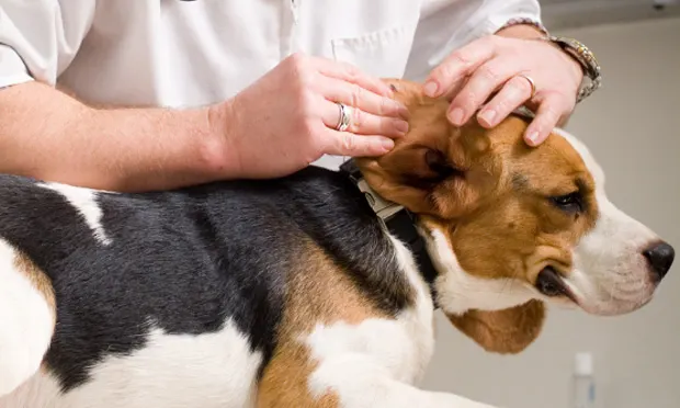
Dysfunction of the vestibular system is a common neurologic disorder. Localization of vestibular system lesions is crucial to generate a differential list and diagnostic plan appropriate for individual patients, as well as to provide owners with a realistic prognosis for their pet. The following cases illustrate some common, and not-so-common, neurologic presentations of animals with vestibular disease.
Related Article: Vestibular Disease
CASE #1
History
Bella, a spayed Yorkshire terrier (4 years of age), presented for progressive balance loss. Her owner had noted stumbling and circling, most notably on stairs and near furniture, for 10 days’ duration. Two years earlier, Bella was diagnosed with immune-mediated hemolytic anemia (IMHA), which was currently in clinical remission with no medications administered. Bella stayed indoors and had no history of travel or toxin exposure. She was not current on vaccinations, as none were given after diagnosis of IMHA.
Physical Examination
Unremarkable with the exception of grade II medial patellar luxations (MPLs) bilaterally
Neurologic Examination
Mentation
Quiet to dull
Cranial nerves
Left-sided head tilt
Resting rotary nystagmus with the fast phase to the left
Positional vertical nystagmus
Positional ventral strabismus in the left eye when head held in extension
Left-sided lip and facial droop
Menace response, pupillary light responses and facial sensation all considered normal.
Gait
Moderate vestibular ataxia—fell and veered to the left, occasionally circled left
General proprioception
Delayed placing response in left pelvic limb, delayed hopping and hemi-walking on left limbs
Spinal reflexes
Normal
Pain response
Winced on palpation of cervical spine and head
Ask Yourself
What is the neurologic anatomic diagnosis?
Can signs be explained by one lesion?
What are the next most appropriate diagnostic tests?
What are the most likely differentials?
Related Article: Motion Sickness in Small Animals: Pathophysiology & Treatment
Lesion Localization
Neurologic examination of Bella suggests a left-sided central vestibular lesion. Her proprioceptive deficits, along with the fast phase of her nystagmus being to the same side as her head tilt, and the nystagmus becoming vertical positionally all indicate a central lesion. The other neurologic examination abnormalities can be found in patients with either central or peripheral vestibular dysfunction and do not help localize the lesion to the brain or periphery.
Differential Diagnosis
Inflammatory disease (infectious or noninfectious/immune-mediated origin, such as granulomatous meningoencephalitis [GME] or necrotizing leukoencephalitis [NLE])
Neoplasia (glioma, meningioma, lymphoma, extension of aural or skull neoplasia)
Malformation (eg, Chiari-like malformation, intracranial arachnoid cyst)
Recommended Diagnostic Tests & Results
CBC/serum biochemistry panel/urinalysis: Unremarkable
Blood pressure: 140 mm Hg on Doppler
MRI of brain: Solitary, indiscrete, intraaxial lesion in the left medulla with mild swelling of the brainstem and nonhomogenous enhancement after IV contrast was administered
CSF analysis from cerebellomedullary cistern: Moderate lymphocytic pleocytosis (105 cells; normal, <5 cells), moderately elevated protein (89 mg/dL; normal, <25 mg/dL)
Outcome
Infectious disease titers were completed, and proved negative, for the following: Toxoplasma gondii, Neospora caninum, distemper virus, Cryptococcus spp, Rickettsia ricketsii, Anaplasma spp, and Ehrlichia canis. A presumptive diagnosis of granulomatous meningoencephalomyelitis (GME) or meningoencephalitis of unknown etiology (MUE) was made.
Oral prednisone was initiated at an antiinflammatory dose of 0.5 mg/kg q12h in addition to modified cyclosporine (Neoral) at 5 mg/kg q12h and cytosine arabinoside (Cytosar) at 100 mg/m2 for 4 doses, q4wks. In the author’s experience, when multimodal forms of immunosuppression are used, immunosuppressive prednisone therapy is rarely needed and precludes dogs from experiencing even more profound prednisone adverse effects. Therapy was tapered based on resolution of neurologic signs, and Bella remained neurologically normal one year later.
CASE #2
History
Oliver, an intact male beagle (6 years of age), presented for waxing and waning ataxia over 2 to 3 weeks and odd head movements. He had no pertinent past medical history and was not on any medications. He was current on his vaccinations and had no history of travel or toxin exposure.
Physical Examination
Unremarkable general physical examination
Neurologic Examination
Mentation
Normal, very bright, and alert
Cranial nerves
Mild head tilt varied at times between right and left
Wide lateral head excursions to both directions
Resting horizontal nystagmus that was fast-phase to the left and did not change phase when head held in extension
Decreased palpebral reflexes bilaterally, left worse than right
Menace responses slightly decreased in both eyes commensurate with decreased palpebral reflex, but vision appeared normal based on elevation of third eyelids and retraction of globes during menace testing.
Pupillary light responses and facial sensation considered normal.
Gait
Nearly normal, but occasionally had a vestibular ataxia with leaning and veering to the left
General proprioception
Normal in all limbs
Spinal reflexes
Normal
Pain response
No pain on palpation of spine or head appreciated
Ask Yourself
What is the neurologic anatomic diagnosis?
Can signs be explained by one lesion?
What are the next most appropriate diagnostic tests?
What are the most likely differentials?
Lesion Localization
Neurologic examination in this case suggested bilateral peripheral vestibular disease and bilateral facial nerve paresis. The head tilt varying to both sides and wide lateral head excursions are typical of this localization. Peripheral vestibular disease also commonly affects cranial nerve VII (facial nerve), as the course of the facial nerve travels in close proximity to the middle ear, so Oliver’s palpebral deficits should not be confused with a central localization. Of note, it is not uncommon for animals with bilateral peripheral vestibular disease to have little or no head tilt and nearly normal gait, as these deficits are much more apparent when one side of the vestibular system is solely or more dramatically affected.
Differential Diagnosis
Bilateral otitis media and/or interna
Endocrine (hypothyroid) related vestibular disease
Bilateral idiopathic vestibular disease with concurrent bilateral facial paresis (less commonly seen)
Recommended Diagnostic Tests & Results
CBC and serum biochemistry panel: Cholesterol of 780 mg/dL, (normal, 92–324 mg/dL) mild non-regenerative anemia of 30% (normal, 36–60%)
Urinalysis: Normal
Blood pressure: 110 mm Hg on Doppler (normal, 90–130 mm Hg)
MRI of brain and tympanic bullae: Normal
CSF analysis from cerebellomedullary cistern: Normal
T4: <0.5 mcg/dL (normal, 0.8–4 mcg/dL)
Free T4 by equilibrium dialysis: <0.5 ng/dL (normal, 0.7–3.7 ng/dL)
Outcome
Oliver was diagnosed with hypothyroid-related neuropathy causing his bilateral vestibular and facial nerve signs. Levothyroxine was initiated at 0.03 mg/kg POq12h and, at recheck four weeks later, Oliver’s vestibular signs had completely resolved, and his bloodwork indicated a euthyroid state (Total T4 3.4 mcg/dL).
CASE #3
History
Marcus, a castrated shih tzu (12 years of age), was presented for peracute onset of severe balance loss and circling for one hour. He had a recent medical history of polyuria and polydipsia, and he was currently treated with enalapril (Enacard) at 0.4 mg/kg PO q24h and a prescription diet for kidney function for mild protein-losing nephropathy diagnosed one year ago. He had no history of travel and was current on vaccinations.
Physical Examination
Pot-bellied appearance
Immature cataracts bilaterally
Crepitus in carpi and left stifle, which also had a grade III MPL
Moderate dental calculus and periodontal disease
Neurologic Examination
Mentation
Quiet and alert
Cranial nerves
Right-sided head tilt
No resting nystagmus noted, but there were occasional beats of positional rotary nystagmus, fast phase to the left
Positional ventral strabismus in the right eye
Absent menace response in the right eye, but appeared visual when walking
Palpebral and pupillary light responses were considered normal.
Gait
Tightly circling to the right, falling to the right
When raising his head on his own, patient would rear up and fall backwards
Coarse body tremor when standing (ie, titubation)
Right limbs hypermetric
General proprioception
Placing responses normal in all limbs
Spastic and delayed hopping responses in right limbs
Spinal reflexes
Normal
Pain response
Winced on palpation of cervical spine and thoracolumbar junction
Ask Yourself
What is the neurologic anatomic diagnosis?
Can signs be explained by one lesion?
What are the next most appropriate diagnostic tests?
What are the most likely differentials for Marcus?
Lesion Localization
Neurologic examination of Marcus suggested a right-sided cerebellar lesion. All vestibular signs can be seen with cerebellar disease. In addition, the coarse body tremor, hypermetric gait, spastic hopping testing, and menace deficit with normal vision would be consistent with a lesion of the cerebellum, and, because all deficits were on the right side of the body, the lesion would be on the right as well.
Differential Diagnosis
Vascular event (ischemic or hemorrhagic infarct)
Inflammatory disease (infectious or noninfectious/immune-mediated origin, such as GME)
Neoplasia (glioma, meningioma, lymphoma, extension of skull neoplasia)
Malformation (intracranial arachnoid cyst)
Recommended Diagnostic Tests & Results
CBC/serum biochemistry panel: BUN 53 mg/dL (normal, 6–31 mg/dL), creatinine 2.1 mg/dL (normal, 0.5–1.6 mg/dL)
Urinalysis: Specific gravity 1.013
Blood pressure: 210 mm Hg on Doppler (normal, 90–130 mm Hg)
Abdominal ultrasound: Chronic degenerative changes to kidneys
Thoracic radiographs: Normal for patient age
MRI of brain: Discrete, wedge-shaped hyperintense lesion of the right cerebellum in the distribution of the rostral cerebellar artery. No mass effect to the lesion and minimal peripheral enhancement after contrast administration indicating vascular compromise or breakdown of the blood-brain-barrier.
CSF analysis from cerebellomedullary cistern: Not performed at owner request
Outcome
Based on the MRI, a nonhemorrhagic infarction of the cerebellum was diagnosed. Given the hypertension, azotemia, and isosthenuria, as well as the dog’s history of PLN, a urine protein-creatinine ratio was repeated and was 6.2 (normal, <0.5). Amlodipine (Norvasc) was initiated at 0.14 mg/kg q24h and his enalapril was increased to 0.8 mg/kg q24h.
Blood pressure was rechecked 4 days later and was 130 mm Hg.
At recheck 3 weeks later, Marcus’s gait and circling had improved vastly, but the head tilt persisted. Recheck UPC showed improvement with a result of 3.0.