Transoral Tracheal & Transtracheal Airway Wash
Elizabeth Rozanski, DVM, DACVECC, DACVIM, Cummings School of Veterinary Medicine at Tufts University
Pulmonary diseases are common in dogs and cats. Although some patients are treated with empirical therapy and recover uneventfully, others require more thorough diagnostics, including sample collection for cytologic analysis, bacterial culture and sensitivity testing, and possible PCR analysis.
Options for Sample Collection
Several options exist for collecting samples from the respiratory system; each has advantages and disadvantages. Standard care for diagnosing pneumonia in humans is sputum culture, but this requires patient cooperation. In rare cases, cultures may be pursued in dogs when sputum production is visualized, but sputum cultures should generally be considered minimally useful for diagnosing pneumonia in dogs.1
Although some dogs and cats with pulmonary disease are treated with empirical therapy and recover uneventfully, others require more thorough diagnostics.
Oral swabs have been minimally evaluated in dogs, with one study showing limited efficacy for isolation of Bordetella bronchiseptica.1 PCR testing has been used with oral swabs, but results should be evaluated in conjunction with clinical signs and careful assessment of laboratory standards.
Fine-needle lung aspiration of focal lesions is used occasionally but is often reserved for cases in which neoplasia is suspected and used less commonly when an infectious process (eg, pneumonia) is suspected. Moreover, while there may be a high diagnostic yield with a pulmonary aspirate, aspiration of diseased lung samples may be associated with such complications as iatrogenic pneumothorax.2
Tracheal Wash
In most cases of suspected lower airway disease, the technique for pulmonary sample collection is a tracheal wash. Tracheal wash samples are useful for airway cytologic evaluation and bacterial culture and sensitivity testing.
The clinician is generally looking for cytologic confirmation of inflammation (neutrophils), infection (degenerative neutrophils with intracellular bacteria), allergy (eosinophils), parasites (larvae), or, rarely, neoplasia in the airway. Bacterial culture results from tracheal wash samples should be interpreted in light of the cytology, as the larger airways are not sterile and simple positive bacterial culture results do not distinguish between airway colonization and infection.
Method Selection
A tracheal wash may be performed via either a transoral tracheal (TOTW) or transtracheal (TTW) approach, depending on clinician preference and patient characteristics. TOTW is performed in briefly anesthetized, intubated dogs, whereas TTW is performed in an awake or lightly sedated patient by inserting a catheter through the trachea into the lower airways. In both approaches, saline is infused to help retrieve airway secretions.
In the author’s experience, TOTW is preferable in small dogs (<10 kg), cats, and brachycephalic dogs because they commonly have a smaller tracheal diameter and thicker necks, which may make tracheal palpation more challenging. TOTW may also be performed in larger dogs, although TTW is preferred in medium- to large-sized dogs. In very large dogs (eg, the Great Dane), the length of the through-the-needle catheter may limit sample collection with TTW; therefore, TOTW using a long catheter may be preferred.
TOTW is technically simple and may be preferred in practices where tracheal wash procedures are performed infrequently. Drawbacks of TOTW include the need for brief general anesthesia sufficient to permit intubation and decrease or eliminate the cough reflex, which may diminish sample quality. Potential complications of TTW include subcutaneous emphysema, tracheal laceration, or hemorrhage.3 Potential complications of saline wash in dogs include hypoxemia and bronchospasm, although these are unlikely in a stable patient.3,4 In any compromised patient, the risk:benefit ratio of performing a tracheal wash should be considered carefully.
Before & After Sample Collection
Before either procedure, the patient should be evaluated for stability. Supplies must be gathered, and an assistant must be on hand.
Recovered fluid should appear cloudy, with occasional flecks of mucus. Cytologic examination (eg, neutrophils and/or intracellular bacteria) should be performed in house immediately after sample collection. Ideally, the sample should then be submitted to a veterinary clinical pathologist for interpretation and aerobic culture and sensitivity testing. The patient should be monitored carefully during recovery.
Step-by-Step: Transoral Tracheal Wash
What You Will Need
Sterile lubricant
IV catheter
Anesthetic agent (typically propofol alone or combined with a low-dose opioid or benzodiazepine)
Supplemental oxygen
Anesthetic machine or bag valve mask (may not be needed)
Monitoring equipment (pulse oximeter, electrocardiograph)
Sterile endotracheal tube (consider using a tube 0.5–1 size smaller than usual)
Sterile gloves and laryngoscope
3 aliquots of sterile saline
Sterile red rubber catheter
Suction device or urine cup
EDTA tubes for sample aliquot for cytology
Plain tube or culturette for bacterial culture

Step 1
Induce brief general anesthesia (typically propofol 4–8 mg/kg IV to effect) to permit intubation. Propofol allows rapid recovery and is helpful if concurrent evaluation of laryngeal function is planned.
Author Insight
Some clinicians prefer to add opioids or benzodiazepines to the anesthetic protocol.

Step 2
Using clean technique, intubate the patient with a sterile endotracheal tube. If sterile lubricant is used, carefully apply a small amount to the outside of the tube.
Author Insight
A slightly smaller-than-normal tube can permit easier intubation. The author prefers not to lubricate the tube because of potential interference with cytologic interpretation and culture results.

Step 3
Pass a 5- to 8-Fr red rubber catheter down the endotracheal tube to approximately the level of the carina (heart base). Alternatively, use a suction catheter or closed suction trap device. Suction devices may permit better recovery of airway cells but are less widely available than red rubber catheters.5 Infuse saline: usually 3 mL for small dogs and cats, 5 mL for medium-size dogs (10–20 kg), and 10–15 mL for large dogs (>20 kg).

Step 4
Manually aspirate fluid. If using a suction device, connect the device directly to a suction unit. Fluid may also be collected directly as it is expectorated from the endotracheal tube into a sterile urine collection cup (shown). Suction additional fluid as needed. Drainage may be facilitated by lifting the patient’s hindquarters.
Author Insight
Before extubation, the patient should be awake enough to facilitate a cough reflex and to prevent complications (eg, reflux, aspiration).

Step-by-Step: Transtracheal Wash
What You Will Need
Local anesthetic (eg, lidocaine)
Clippers
Surgical preparation solution
Through-the-needle catheter (preferred) or needle and polypropylene or red rubber catheter (small enough to feed through the needle)
3 aliquots of sterile saline

Step 1
Manually restrain the dog on the floor or a treatment table. Clip and prepare a site over either the lower cervical trachea or the cricothyroid ligament. After carefully palpating the intended catheter placement site, infuse a local anesthetic such as lidocaine (not shown). It is often easier to isolate the cricothyroid ligament in dogs with thicker necks (eg, Labrador retrievers).
Author Insight
Restraining the dog in a corner may help prevent efforts to escape.

Step 2
With the bevel directed downward, insert the needle through the skin and into the tracheal lumen. The needle will typically be perpendicular to the trachea.

Step 3
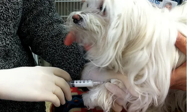
Advance the catheter through the needle down the trachea. Pull the needle from the skin, and place a needle guard to avoid inadvertent laceration of the catheter. Remove the stylet. When the catheter is successfully placed within the tracheal lumen, air can be aspirated easily from the catheter; the dog will usually cough.
Author Insight
Some clinicians protect the needle manually rather than placing a needle guard; however, this step is important to avoid damaging the catheter.
Step 4
Infuse saline (5- to 10-mL aliquots), then aspirate to collect the sample. Repeat up to three times. Gentle coupage may help encourage coughing, which can enhance sample quality.
TOTW = transoral tracheal wash, TTW = transtracheal wash