Top 5 Tips for Improving Dental Imaging
Sponsored by iM3
Obtaining high-quality dental radiographs is an essential component of companion animal medicine. In 2 studies that examined the impacts of dental radiography on dogs and cats, intraoral radiography was shown to reveal clinically significant pathology in 27.8% of dogs and 41.7% of cats.1,2 Without dental radiography, oral pathology can go undetected and untreated, impacting patient health and quality of life.
Although high-quality dental radiographs are essential, capturing quality images can be a challenging and time-consuming task for the veterinary team. Obtaining these images requires a well-trained team and optimal equipment, and dental radiography involves a steep learning curve for many veterinary professionals.
These 5 tips may help to alleviate some of the stress associated with dental radiography.
1. Use a Veterinary-Specific Dental X-Ray Generator
Although some veterinarians may operate with human dental equipment in their practice, purchasing veterinary-specific equipment that has been reviewed and approved by the FDA for veterinary use can provide numerous benefits.3 Veterinary specific x-ray generators offer greater usability and suitability for canine and feline patients and an improved experience for the veterinary team capturing images.
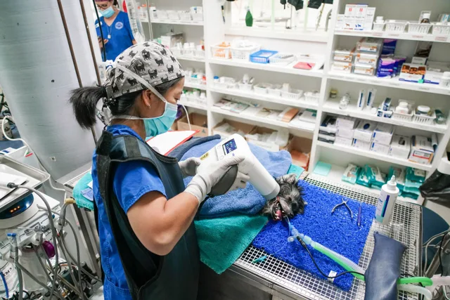
Dental x-ray generators are available in various forms to meet a variety of practice needs. Larger dental x-ray generators, such as the iM3 Revolution 4DC, are available as wall-mounted units or on a wheeled trolley. The imaging head of these generators can be positioned and locked in place, allowing the veterinary team to take radiographs from a distance using a handheld wireless remote. In smaller spaces, portable, handheld dental x-ray generators, such as the iM3 Port-X IV, can provide flexibility. The iM3 Port-X IV also includes a back scatter shield, minimizing radiation exposure when the operator must remain close to the patient.
2. Select a User-Friendly Software System
A variety of software solutions are available for digital veterinary dentistry, but it is important to select software that is user-friendly, as this can help promote practice and team efficiency. This will reduce the time spent taking and viewing radiographs, allowing more time to be spent working directly with patients.
The user-friendly iM3 Vet Exam Pro includes veterinary-specific layouts that are appropriate for a large range of species. This software allows for easy comparison between new images and saved images from the same patient while also allowing for viewing 6 to 7 images in a single layout.
3. Adopt a Systematic Approach to Dental Radiography
When taking full-mouth radiographs, a systematic approach (ie, taking the same views in the same order on every patient) can help ensure that nothing is missed. Software layouts, such as those included in the iM3 Vet Exam Pro, can help team members acquire these images quickly and efficiently.
A systematic approach should also be used when interpreting radiographs to help ensure that all captured structures are evaluated for potential pathology.
4. Use Positioning Aids to Improve Image Quality
Obtaining diagnostic dental radiographs requires careful positioning to visualize all structures of the tooth. This requires an understanding of bisecting angles to minimize image distortion and ensure that all relevant structures are visible.
The iM3 X-Ray Positioning Kit aids in positioning, providing preset guides to help achieve the correct relative angle, distance, and position for diagnostic dental images. This kit is designed to provide full-mouth radiographs in just 6 radiographic views. Using this kit can provide a simple, reproducible method that allows any veterinary professional to obtain diagnostic dental radiographs.
5. Have a Variety of Plate Sizes Available to Accommodate a Range of Patients
Taking the best possible dental radiographs requires access to a variety of plate sizes to accommodate all patient sizes. When using iM3 plates, sizes range from 20 mm × 30 mm (size 0) to 50 mm × 140 mm (size 6). To be prepared for patients of any size, clinics should ensure they have a range of sizes available.
Conclusion
Dental radiography is an essential component of dental care in companion animal patients. Appropriate training and optimal dental equipment can increase both the quality of dental radiographs and the ease and efficiency with which radiographs are taken.