Diet-Related Canine Skin Conditions
Susan Paterson, VetMB, MA, DVD, DECVD, FRCVS, Virtual Vet Derms, Kendal, United Kingdom

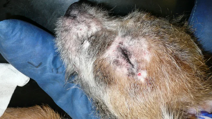
Crusted lesions on the face of a husky with zinc-responsive dermatosis
YOU HAVE ASKED...
How can diet cause skin disease in dogs?
THE EXPERT SAYS...
Skin disease can be associated with diet via nutritional deficiencies (inherited or acquired) or adverse reactions (immunologic or allergic).
Nutritional Deficiency
Acquired nutritional deficiencies are rare because of the quality and regulation of commercial diets. Nutritional imbalances—most commonly related to zinc, vitamin A, fatty acids, and protein1—more often occur when dogs with specific nutritional requirements (eg, because of age or disease risk) are fed inappropriate diets or when systemically poor health affects a dog’s ability to use specific nutrients.
Zinc
In dogs, 2 manifestations of zinc-responsive dermatosis have been documented.2,3
The first, caused by a genetic inability to adequately absorb zinc, most commonly occurs in young adult dogs; affected dogs are usually fed balanced diets. Northern breeds (eg, Siberian husky, Alaskan malamute, Samoyed) seem predisposed (Figure 1). Affected dogs are presented with variable degrees of pruritus, crusting, alopecia, and erythema. Lesions are usually located over pressure points as well as the periocular and perioral area, pinnae, pads, and planum nasale. Secondary pyoderma is common. Affected dogs need lifelong zinc supplementation.3-5
The second occurs in young dogs fed a diet either deficient in zinc or with high levels of ingredients that reduce zinc bioavailability. Food abundant in plant phytates, calcium, cereal, or soy can interfere with zinc absorption in the GI tract. Affected dogs are typically presented with a crusting dermatosis of the mucocutaneous junctions, pressure points, and trunk. Lesions may resolve with a balanced diet.5,6
Idiosyncratic reactions to food can mimic drug reactions and can manifest with a wide array of clinical signs.
Vitamin A
Vitamin A is important for maintaining healthy skin and epithelial cells. Deficiency and toxicity cause similar signs: epidermal hyperkeratosis and scaling, poor hair coat, and alopecia.7 Because of its function in maintaining healthy skin, vitamin A has been used to treat severe seborrhea in some breeds.8
Polyunsaturated Fatty Acids
Essential for growth and reproduction, polyunsaturated fatty acids (PUFAs) are also vital for preventing skin lesions. Linoleic acid and linolenic acid have long been recognized as essential PUFAs in dogs. Fatty-acid deficiency is usually present for several months before cutaneous signs become evident.7,9 Dogs develop fine scaling and loss of hair luster, often with alopecia and bacterial pyoderma. In chronic cases, the skin can become thickened with a greasy seborrhea; secondary yeast infections are common.
Fatty-acid deficiency is rare but has been recorded in dogs fed dry rations, poorly stored commercial food, homemade rations, or poorly formulated low-calorie diets.7 PUFA deficiency can be caused by food oxidation from prolonged storage (canned food, 1 year; dry food, 6 months), inadequate antioxidants, or high temperature. Fatty-acid deficiency can also occur when the diet is nutritionally complete but PUFA absorption is reduced because of intestinal malabsorption or exocrine pancreatic insufficiency. Impaired PUFA biosynthesis due to chronic hepatic disease can lead to similar signs.10
Protein
Protein deficiency (rare) is usually associated with starvation or low-protein diet. Affected dogs may have hyperkeratosis, epidermal hyperpigmentation, and loss of hair pigment. Hair growth puts huge protein demands on the body, so protein deficiency can affect hair growth and cause alopecia as the hair becomes thin, rough, dry, dull, and brittle.10
Adverse Food Reactions
Adverse food reactions (ie, reactions to dietary allergens) can be classified as food allergy or food intolerance. Food allergy is an immunologically based reaction, which may involve both type-I and type-III immune reactions. Food intolerance includes metabolic, pharmacologic, and idiosyncratic reactions as well as intoxication (eg, from bacterial and fungal toxins).11 (See Case Study: Idiosyncratic Food Reaction.)
Cutaneous adverse food reactions (CAFRs) likely play an important part in canine atopic dermatitis.13 Current thinking supports the idea that CAFR might manifest as atopic dermatitis in some dogs; however, dogs with CAFR may also experience clinical signs (eg, GI signs) not typically associated with atopic dermatitis. It has been suggested that atopic dermatitis be divided into food-induced atopic dermatitis and nonfood-induced atopic dermatitis—or canine atopic dermatitis sensu stricto for cases not responsive to elimination diets. Numerous studies have been published on dermatologic manifestations of CAFR in dogs.11,14-20
Self-inflicted trauma to the face of a young border terrier with CAFR.
Most CAFR cases occur in young dogs. Pruritus caused by CAFR is more common than atopic dermatitis in dogs <6 months of age.11 Nonseasonal pruritus, a consistent finding,11,16,20 is often poorly responsive to glucocorticoids.20 Cutaneous signs of CAFR in the dog overlap with other allergic dermatoses involving facial, pinnal, and ventral skin.16 Pruritus of the ears and licking of the perianal area are common,11 but CAFR can affect only the perianal skin.21 When present, primary lesions are usually papular; secondary lesions in the form of excoriation from self-inflicted trauma (Figure 2) can be complicated by bacterial or yeast infection.
The gold standard for CAFR diagnosis is abatement of signs while the patient is fed an appropriate restricted or novel diet and recurrence of signs when the patient is challenged with previous food items. Identifying a truly novel protein diet can be difficult, and the availability of hydrolyzed diet over the past 10 years has led to the institution of more effective diet trials.22-25 Other tests (eg, intradermal skin testing,26,27 skin patch testing,28 measuring circulating food allergen–specific serum IgE26,29) are of no diagnostic value because of low sensitivity and specificity.
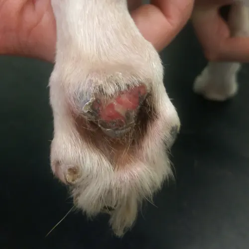
Punched-out ulcers on the pad of a dog with vasculitis.
CAFRs may present as vasculitis (eg, punched-out ulcers in the center of pads [Figure 3], ulceration and crusting of the pinnal margin, ulcerated lesions on the concave aspect of the pinna), and urticarial vasculitis.30,31 The lesions of urticarial vasculitis present with urticaria-like lesions; unlike true urticaria, however, they do not blanche with diascopy and do not pit with pressure.30
Case Study: Idiosyncratic Food Reaction
In addition to cutaneous adverse food reactions, idiosyncratic reactions to food can mimic drug reactions and can manifest with a wide array of clinical signs. One study describes a border collie presented with erythematous lesions in the axillae, groin, mucocutaneous junctions, and pinnae. Erythema multiforme was diagnosed on histopathology. Lesions responded to azathioprine, prednisolone, and a hypoallergenic diet. The disease did not relapse as drugs were withdrawn but recurred every time the original commercial diet was reintroduced; this suggests the diet caused the disease.12