Determining Canine Estrus Stage via Vaginal Cytology
Autumn P. Davidson, DVM, MS, DACVIM, University of California, Davis

Performing vaginal cytology offers a rapid, inexpensive, and reliable in-clinic method to evaluate stages of the estrus cycle in the bitch. Veterinary discomfort with obtaining and interpreting vaginal cytology is common; submission to a commercial laboratory might result in diagnostic delays and increased client costs.
Equipment required for vaginal cytology (cotton-tipped applicators, frosted microscope slides, commercial Romanowsky [Diff-Quik] stain, and light microscope) is already present in most small animal practices.
Competence in vaginal cytology allows a clinician to:
Determine whether a bitch is actually in heat.
Aid in determining the correct time to begin performing more expensive serum progesterone and luteinizing hormone (LH) assays for precise ovulation timing.
Determine if it is too late in the estrus cycle to perform artificial insemination in dogs unable or unwilling to breed naturally.
Determine if a bitch is under the influence of estrogen (endogenous or exogenous).
Predict the correct day to perform an elective cesarean section (C-section).
Proper Technique
Proper technique is important so that cells obtained are representative of hormonal changes. The sample should be collected from the cranial vagina because cells from the clitoral fossa, vestibule, urethral papilla, or vestibulovaginal junction are not as indicative of the stage of the cycle and provide confusing results (A). A cotton-tipped applicator (moistened with water if needed) should be passed into the vulva in a dorsal direction and advanced horizontally above the clitoral fossa and urethral papilla into the vagina, which is at the level of the cranial thigh (B).


After swabbing (by gently rubbing or rolling) the vaginal wall, the applicator is removed and rolled (not smeared) onto a glass slide (C). The slide should be labeled, including name and date (with a pencil).

Routine Diff-Quik staining is performed after air-drying the slide. Scan the slide at low power first (10×) and high power as necessary (40×) to aid in particular cell identification. It is best to survey a large area of the slide for cell types.
Vaginal Cytology
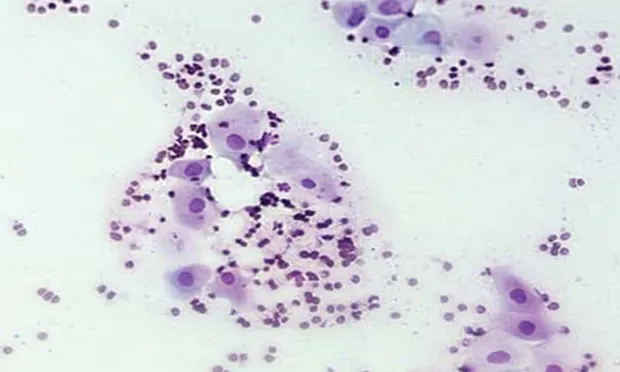
Six types of cells typify vaginal cytology: parabasal cells (A), which look like small, O-shaped oat cereal pieces; small intermediate and large intermediate cells (B), which look like fried eggs; superficial (“cornified”) (C) cells, which look like corn flakes; neutrophils; and red blood cells.

Remember the Estrus Cycle
The 4 phases of the estrus cycle are:
Anestrus (not in heat): Parabasal cells predominate; the cellularity is low (A).
Proestrus (early estrogen influence): From early to late proestrus, a gradual shift from parabasal and intermediate cells (small then larger) and finally superficial cells occurs (B). Typically, red blood cells are present in large numbers (C).
Estrus (receptive, fertile): Superficial cells predominate and their nuclei become pyknotic or absent/anuclear (D, E).
Diestrus (the luteal phase): Onset of diestrus is marked by a precipitous decline in the number of superficial cells and reappearance of intermediate and parabasal cells within 1 to 2 days. Neutrophils are commonly observed (F), and large numbers of bacteria are also often present (G).

1. Anestrus (not in heat):
Parabasal cells predominate; the cellularity is low (A).
Questions Answered Through Vaginal Cytology
Is the bitch in heat? As estrogen rises during proestrus, maturation rate of the vaginal epithelial cells increases, as does the number of keratinized, cornified epithelial (ie, superficial) cells seen on a vaginal smear. Full cornification continues throughout estrus, which prepares the vagina for natural breeding. Proestrus and diestrus cytologies can be similar (parabasal and intermediate cells ± neutrophils). Rechecking in 48 hours can clarify the stage.
When should progesterone be tested for ovulation timing? Breeder clients should be advised to notify the clinic when they first notice vulvar swelling, vaginal discharge, or attraction to males in a bitch for which breeding is planned. Early proestrus should be documented by vaginal cytology (<50% cornified [superficial cells]). Vaginal cytology should be performed every 2 to 4 days until a significant progression in cornification is seen, usually above 70% superficial cells. At that point, serial hormonal assaying should begin.
Is it too late to breed? At the end of the fertile period, vaginal cytology undergoes the diestrual shift, which signifies the first day of diestrus. Breeding after the diestrual shift is rarely successful.
Is the bitch under the influence of estrogen (eg, ovarian remnant, unspayed, exposure to owner’s hormone replacement medication)? The defining characteristic of estrogen influence is the presence of superficial cells in vaginal cytology, but this should be confirmed with additional tests.
How do I predict the right day for an elective C-section? If vaginal cytology is performed until the diestrual shift is observed, a retrospective analysis of the date of the LH surge (7–10 days previously), ovulation and ova maturation (approximately 24–48 hours after the LH surge), and the fertile period (approximately 3–6 days after the LH surge) can be obtained. It is the least expensive way to perform ovulation timing, albeit retrospectiveIy, and can be useful if evaluation of gestational age becomes important, as parturition (C-section) should occur 56 to 58 days from the day of the diestrual shift.