To Cut or Not to Cut: Toe Lesion in a Cat
Signalment
Spayed domestic shorthair cat of unknown age.
History
Adopted as an adult 5 years earlier. Indoor only cat. Limping on right pelvic limb for approximately two weeks.
Physical Examination
Grade IV/IV right pelvic limb lameness. Very painful, palpable soft tissue swelling of the third digit. No detectable discharge or wound along the toe.
Radiographic Findings
One dorsal–plantar view of the tarsus and foot of the right pelvic limb was obtained (Figure 1). A lateral view was not obtained because of the likelihood of low diagnostic yield (ie, superimposition of digits 2–4). The toes can be separated with gauze or string for a lateral view, but this patient was too painful to do so without analgesia/sedation.

Soft tissue swelling surrounded the phalanges of the right pelvic limb third digit. The entire distal phalanx of the third digit was absent, and the distal portion of the middle phalanx of the third digit was lytic.
Diagnosis
Aggressive bone lesion of the distal and middle phalanges of the third digit. Aggressive bone lesions can be secondary to neoplasia or infection. Absence of periosteal proliferation and the degree of lysis makes neoplasia more likely than infection. The involvement of two adjacent bones also suggests a soft tissue origin of malignancy (primary bone tumors rarely cross joints). Metastasis of pulmonary carcinoma to the digit is well documented1-3 and should be a strong differential at this point.
Should This Cat Go to Surgery?
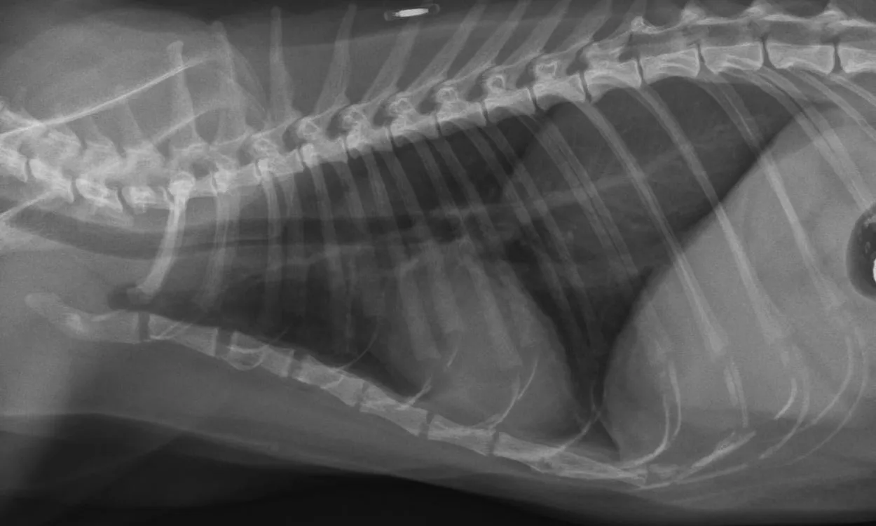
Thoracic radiographs (Figures 2A and 2B) were obtained to check for primary or metastatic neoplasia; an approximately 8.3 cm long, 3.8 cm tall, and 3.6 cm wide soft-tissue mass was present in the dorsal aspect of the right caudal thorax (white arrows), occupying the majority of the right caudal lung lobe. The aortic arch is prominent in the ventrodorsal view (black *); this is common in geriatric felines and may be a variation of normal or possibly an indication of systemic hypertension. The finding of a mass in the lung of this cat supports a diagnosis of a primary pulmonary carcinoma originating from the right caudal lung lobe with metastasis to the third digit of the right pelvic limb. This syndrome is described in several case reports.1-3 Most cats have swelling of one or more digits with radiographic evidence of osteolysis of the affected digit(s). The tendency of feline pulmonary carcinomas to metastasize to one or more digits is not understood, and no specific treatment has been rewarding.


Outcome
The patient received buprenorphine transmucosally q8h for pain management, but ultimately succumbed to pulmonary neoplasia with intrathoracic metastases and severe pleural effusion. The lung and digit were submitted for histopathology; papillary carcinoma of the lung and metastatic pulmonary carcinoma of the digit were confirmed.