The Case: Remnant Urethral Calculus After Cystotomy

Clinical History
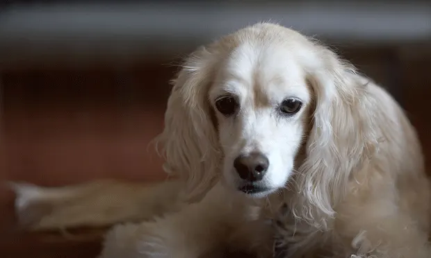
6-year-old spayed female Cocker spaniel mix with stranguria, hematuria, and pollakiuria of 5 days’ duration.
Physical examination revealed normal-sized urinary bladder. Remainder of physical examination was within normal limits.
CBC/serum biochemical profile: within normal limits
Urinalysis- specific gravity: 1.040- pH: 7.0- 2+ blood, 2+ protein, 3–5 RBCs/hpf- 5–10 WBCs/hpf- 5–10 calcium oxalate crystals/hpf
Clavulanate-amoxicillin instituted at 13.75 mg/kg Q 12 H.
Survey abdominal radiographs:- several round opaque cystic calculi in bladder- 1 calculus in pelvic urethra- Kidneys/ureters free of calculi
Urine culture/susceptibility: 100,000 CFU Escherichia coli/mL- MIC to clavulanate-amoxicillin: S < 0.5
Owner elected cystotomy/stone removal.
Anesthesia- Premedication: hydromorphone (0.05 mg/kg IM) and midazolam (0.2 mg/kg IM)- Induction: propofol (40 mg IV) to effect- Maintenance: isoflurane inhalant
Surgery- Exploratory laparotomy performed; incision just cranial to umbilicus continuing halfway between umbilicus and pubis.- Bladder exteriorized/ventral cystotomy performed; incision centered near cranial aspect (apex) of bladder dome.- Five calculi removed; bladder closed with double inverting layer using a Cushing suture pattern with 3-0 PDS. Care was taken to avoid entering lumen of bladder with sutures.
Uneventful recovery from anesthesia; patient discharged next morning with carprofen (2 mg/kg PO Q 12 H) and clavulanate-amoxicillin as previously prescribed.
Owners instructed to manage the incision and monitor dog’s urination.
Owner called veterinarian the following day: dog straining repeatedly/unable to pass urine for ~14 hours with signs of abdominal discomfort. Veterinarian referred case to a specialty referral center.
Physical Examination Findings
Patient was depressed; 5% to 7% dehydration
Severely distended, painful abdomen
Diagnostic Procedures
CBC: within normal limits
Serum biochemical profile abnormalities:- BUN 90 (N 6–31 mg/dL)- Cr 4.9 (N 0.05–1.6 mg/dL)- K+ 8.1 (3.6–5.5 mEq/L)
Abdominocentesis: 40 cc of yellow fluid removed- Fluid BUN: 94 (N 6–31 mg/dL)- Fluid Cr: 8.2 (N 0.05–1.6 mg/dL)
Abdominal radiographs:- 1 cystic calculus- Urethral calculus lodged in pelvic urethra- Evidence of peritoneal effusion
ECG: prolonged PR interval/absence of P wave (likely due to hyperkalemia)
Working diagnosis: uroabdomen secondary to remnant urethral calculus
Therapeutic Procedures
Supportive care in ICU:- IV crystalloid fluids- 10% dextrose bolus (4 mL/kg IV), administered twice- Acid–base/serum electrolyte status regularly monitored; metabolic abnormalities corrected within 3 hours. Cardiac abnormalities were reversed as well.
Foley catheter placed in urethra past the calculus and into the bladder; maintained to divert urine away from urinary bladder.
Percutaneous peritonostomy drain placed using local anesthetic and a red rubber feeding tube. Tube placed just off midline (to avoid falciform ligament) at level of umbilicus, allowing efficient drainage of urine from abdominal cavity.
Patient was clinically improved after 6 hours; exploratory laparotomy could now be safely considered as a definitive treatment for the uroabdomen.
Abdominal exploratory procedure:- Anesthetic protocol identical to that of first procedure.- Incision was extended from caudal aspect of original incision to level of pubis. Balfour self-retaining retractors used to maintain retraction of abdominal wall, allowing a complete abdominal exploratory.- Urine suctioned from the abdominal cavity, urinary bladder exteriorized, original bladder-wall incision reopened and extended to bladder neck to facilitate removal of remnant urethral calculus.- One calculus was removed from the bladder, 1 from the urethra, and 1 was found free floating in the peritoneal cavity.- Bladder lavaged to ensure all calculi were removed; piece of mucosa removed for culture/sensitivity.- Bladder closed with a single layer, simple continuous pattern using monofilament synthetic absorbable suture with a swaged-on taper needle. Sutures were placed full thickness in the bladder wall.
Recovery was uneventful; patient discharged the following day.
Clinical OutcomeAt 3 months after discharge, the patient is urinating normally and has had no urinary signs.
The Generalist's Opinion
Nonsurgical options for stone removal include diet therapy for dissolution, voiding urohydropropulsion, electrohydraulic shock-wave lithotripsy, and extracorporeal shock-wave lithotripsy. Given the presence of calcium oxalate crystals, it was reasonable to assume that stone dissolution was not a good option. Lithotripsy carries an extremely high cost; it is not a commonly-performed procedure in veterinary medicine nor is it a realistic option in private practice. Voiding urohydropropulsion is limited to very particular cases in which the stones appear small enough to pass through the urethra and do not have sharp edges that could damage the urethral wall. Thus, the dog in this case had an appropriate surgical cystotomy for the uroliths in its bladder and pelvic urethra.
Attention to Details
Cystotomy can be a fairly straightforward procedure, but if some details are overlooked, it can lead to complications. Studies have shown that uroliths have been incompletely removed in up to 20% of surgical cases.1 Radiographs need to be taken just before surgery to identify any stone that may have shifted position since the diagnosis was made. In addition, a postoperative radiograph must be taken to ensure that all uroliths have been removed.
The first step in the procedure is to remove all identifiable stones either manually, with suction, or with a bladder spoon. Intraoperatively a catheter should be passed into the urethra to flush it thoroughly. Retrograde hydropropulsion can also be used to create negative pressure within the bladder lumen and thus draw uroliths into it. If the need arises, an assistant can pass a catheter externally and flush the urethra in a retrograde direction.
Double-check Your Work
Whatever technique is used, it is important to ensure complete urolith removal and urethral patency prior to closure. If all attempts fail to remove urethral calculi, then an incision into the urethral lumen is required. Options for urethral surgery include an urethrotomy or urethrostomy, both of which require operator experience and familiarity with the particular surgical technique. It is important to recognize that certain cystotomy cases can be far more challenging than others and may require a comfort level with such advanced surgical techniques.
Barak Benaryeh, DVM, DABVP, is the owner of Spicewood Springs Animal Hospital. He graduated from the University of California-Davis School of Veterinary Medicine in 1997 and completed an internship in Small Animal Medicine, Surgery and Emergency at the University of Pennsylvania. Dr. Benaryeh has also taught practical coursework to first-year veterinary students and was a primary veterinary surgeon for the Helping Hands Program, which trains assistance monkeys for quadriplegic people. Dr. Benaryeh is board certified by the American Board of Veterinary Practitioners in Canine and Feline Practice.
Howard B. Seim, III, DVM, DACVS, is a professor of surgery in the Department of Clinical Sciences at Colorado State University College of Veterinary Medicine and Biomedical Sciences. Dr. Seim completed his internship in Saskatoon, Canada, and his residency at the Animal Medical Center in New York. Dr. Seim has been actively involved as an advisor to many veterinary students, interns, and residents, and is a recipient of multiple teaching awards. He is a current member of the NAVC Clinician’s Brief editorial Advisory Board. Dr. Seim is the founder of VideoVet, which provides veterinary surgical continuing education courses on DVD to practicing veterinarians.