In the Literature
Simpson SE, Zersen KM. Incidence and type of peripheral intravenous catheter complications documented in hospitalised dogs. J Small Anim Pract. 2023;64(3):130-135. doi:10.1111/jsap.13574
The Research …
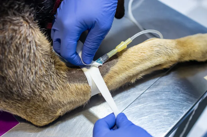
Maintaining patency of IV catheters in hospitalized patients can be challenging. Loss of patency or complications associated with these devices can lead to patient discomfort, catheter-related bacteremia, phlebitis, vascular thrombosis, and increased costs.1-3
This prospective observational study described the incidence of peripheral IV catheter (PIVC) complications (ie, extravasation, phlebitis, dislodgement, occlusion, line breakage) in patients hospitalized in a critical care unit (CCU) and intermediate care unit (IMCU) of a referral teaching hospital and sought to identify risk factors that had a significant effect on PIVC complications. Clinical phlebitis was defined as redness, swelling, pain, or oozing at the PIVC insertion site and was graded using the human visual infusion phlebitis scale. Culture of a catheter was submitted if clinical signs of a fever, redness, pain, heat, or discharge were present. Dislodgement was defined as complete removal of a catheter, whereas line breakage was separation of the T-port line from the male or female adaptor port.
Incidence of PIVC complications in the CCU was 24.2%, with phlebitis being the most common complication. In the IMCU, incidence of PIVC complications was 13.1%, and line breakage was the most common complication. Longer hospitalization and increased patient weight significantly increased the likelihood of PIVC complications. Overall incidence of PIVC complications was 19.9%, and odds for complications were not significantly higher in CCU patients compared with IMCU patients.
… The Takeaways
Key pearls to put into practice:
Although a standardized method of detecting clinical phlebitis has not been determined in human or veterinary medicine, monitoring for redness, swelling, pain, and/or discharge at catheter insertion sites in patients with prolonged hospitalization and implementation of daily catheter management techniques to reduce the incidence of phlebitis are needed.4,5 Standardized preparation and aseptic technique should be used in conjunction with daily assessment for evidence of clinical phlebitis.
Monitoring for PIVC complications and identifying potential risk factors in different patient populations are critical. This study identified hospitalization length and patient weight as risk factors for PIVC complications, but more risk factors may exist.
Routine replacement of PIVCs is not necessary (even in cases of expected prolonged hospitalization), but PIVCs should be removed based on clinical criteria of phlebitis.6
You are reading 2-Minute Takeaways, a research summary resource proudly presented by Clinician’s Brief. Clinician’s Brief does not conduct primary research.